Showing posts with label Surgery. Show all posts

Wednesday, July 12, 2017

Keloid Scars - A Brief Discussion


Keloid scars are tumor like lesions that arise from the connective tissue of a scar and extend beyond the dimensions of the original wound

Predisposing factors

  • ethnicity: more common in people with dark skin
  • occur more commonly in young adults, rare in the elderly
Pathophysiology: When skin is injured, fibrous tissue, called scar tissue, forms over the wound to repair and protect the injury. In some cases, scar tissue grows excessively, forming smooth, hard growths called keloids. Keloids can be much larger than the original wound.


Common Sites (in order of decreasing frequency):

  • sternum, 
  • shoulder,
  • neck,
  • face, 
  • extensor surface of limbs, 
  • trunk
Keloids should not be confused with hypertrophic scars, which are raised scars that do not grow beyond the boundaries of the original wound.

Monday, June 19, 2017

Ruptured Liver Abscess.

A 50-year-old male presented to the Emergency Room with shock and a four-day history of a febrile illness. He required intubation and was started on inotropes.
His Chest X ray is shown below:


Case Discussion: It is important to look at the “blind areas” of the Chest X ray in order not to miss important clues. These areas are under the diaphragm, behind the heart, the hilum, and the soft tissues. This Chest X ray shows a lucency over the liver density. The lucency does not conform to the usual bowel configuration. In this clinical context, an important differential diagnosis to be considered is a ruptured liver abscess. This can be confirmed either by bedside ultrasound or CT,

A CT scan was done in this patient which is shown below:

Wednesday, May 24, 2017

Introduction to Folliculitis, furuncles, and carbuncles

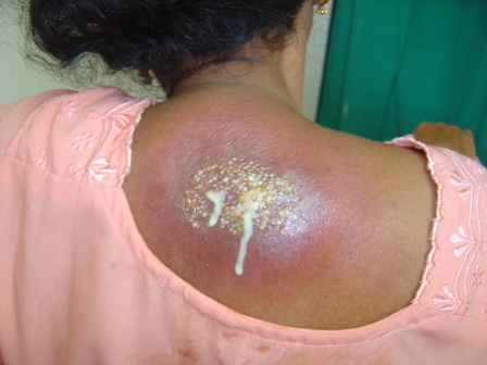
Folliculitis: A bacterial infection of the hair follicle, folliculitis causes the formation of a pustule of the hair follicle opening. The infection can be superficial (follicular impetigo or Bockhart’s impetigo) or deep (sycosis barbae).


Furuncles, commonly known as boils, are another form of deep folliculitis. 


Carbuncles are a group of interconnected furuncles. 

The prognosis depends on the severity of the infection and the patient’s physical condition and ability to resist infection.

Causes

The most common cause of folliculitis, furuncles, or carbuncles is coagulasepositive Staphylococcus aureus. 

Predisposing factors include 
  • an infected wound, 
  • moisture, 
  • obesity, 
  • diabetes mellitus, 
  • skin disease, 
  • poor hygiene, 
  • debilitation, 
  • tight clothes, 
  • friction, and 
  • immunosuppressive therapy.
Signs and symptoms
Folliculitis, furuncles, and carbuncles have different signs and symptoms.

Sunday, May 21, 2017

Intussusception - Clinical Features And Management


With intussusception, a portion of the bowel telescopes (invaginates) into an adjacent distal portion. Intussusception may be fatal, especially if treatment is delayed for a strangulated intestine.

Causes
Intussusception is most common in infants and is three times more common in males than in females. It typically occurs between ages 3 months and 3 years, with a peak incidence between ages 6 and 9 months.
Studies suggest that intussusception may be linked to viral infections because seasonal peaks are noted—in late spring and early summer, coinciding with the peak incidence of enteritis, and in midwinter, coinciding with the peak incidence of respiratory tract infections.
The cause of most cases of intussusception in infants is unknown. In older children, polyps, alterations in intestinal motility, hemangioma, lymphosarcoma, lymphoid hyperplasia, or Meckel’s diverticulum may trigger the process. In adults, intussusception usually results from benign or malignant tumors (65% of patients). It may also result from polyps, Meckel’s diverticulum, gastroenterostomy with herniation, or an appendiceal stump. In the elderly, decreased GI elasticity and motility can lead to intussusception.

Pathology
When a bowel segment (the intussusceptum) invaginates, peristalsis propels it along the bowel, pulling more bowel along with it; the receiving segment is the intussuscipiens. This invagination produces edema, hemorrhage from venous engorgement, incarceration, and obstruction. If treatment is delayed for longer than 24 hours, strangulation of the intestine usually occurs, with gangrene, shock, and perforation.

Sunday, May 14, 2017

Fracture of Nose


Introduction
The most common facial fracture, a fractured nose usually results from blunt injury and is commonly associated with other facial fractures. The severity of the fracture depends on the direction, force, and type of the blow.
A severe, comminuted fracture may cause extreme swelling or bleeding that may jeopardize the airway and require tracheotomy during early treatment. Inadequate or delayed treatment may cause permanent nasal displacement, septal deviation, and obstruction.

Causes
With low-energy injuries, noncomminuted nasal bone fragments are caused by low-velocity trauma. Such injuries could occur in the following situations:
  • injuries created during fistfights (hand or fist blows only, no blunt instruments)
  • uncomplicated falls such as tripping
  • low-velocity motor vehicle collision.
With high-energy injuries, a higher amount of energy is absorbed by the nasal and facial bones, with comminution of bone fragments and associated injuries to the soft tissue and orbitonasal skeleton. These injuries would include:
  • injuries sustained from a leveraged blow to the nose using an object such as a stick, pipe, or other blunt object
  • falls from heights
  • sport injuries with fast-moving projectiles, such as a ball or puck
  • high-velocity motor vehicle collisions.
Clinical Features
Immediately after the injury, a nosebleed may occur, and soft-tissue swelling may quickly obscure the break. After several hours, pain, periorbital ecchymoses, and nasal displacement and deformity are prominent. A possible complication is septal hematoma, which may lead to abscess formation, resulting in vascular septic necrosis and saddle nose deformity.

Thursday, May 11, 2017

Post-traumatic Pneumothorax.

A 24 years old male was brought to radiology department for X-ray chest following a road traffic accident. The X ray is shown below:


Radiological Findings: Chest X-ray shows presence of the air within the right pleural cavity with volume loss of right lung, as a result the right lung has partially collapsed. The outline of collapsed lung is seen well against the air.

Diagnosis: Post-traumatic Pneumothorax.

Clinical Discussion: Presence of the air within the pleural cavity is termed as the pneumothorax.
Air enters into the pleural cavity through the defect in pleural layers either spontaneously or due to trauma.

Wednesday, May 3, 2017

Corrosive Esophagitis

A 50 year old woman was seen in emergency after ingesting a chemical substance . Esophagogastrodudenoscopy was performed and the picture is shown below.


Initial esophagoscopy. (A) Middle esophagus shows whitish discoloration. (B) Distal esophagus shows exudates with easy touch bleeding.

The case was diagnosed as Corrosive Esophagitis 

Corrosive Esophagitis Case Discussion

Introduction
Inflammation and damage to the esophagus after ingestion of a caustic chemical is called corrosive or caustic esophagitis. Similar to a burn, this injury may be temporary or lead to permanent stricture (narrowing or stenosis) of the esophagus that requires corrective surgery.
Severe injury can quickly lead to esophageal perforation, mediastinitis, and death from infection, shock, and massive hemorrhage (due to aortic perforation).
Causes
The most common chemical injury to the esophagus follows the ingestion of lye or other strong alkalies; less commonly, injury follows the ingestion of strong acids. The type and amount of chemical ingested determine the severity and location of the damage.
In children, household chemical ingestion is accidental; in adults, it’s usually a suicide attempt or gesture. The chemical may damage only the mucosa or submucosa, or it may damage all layers of the esophagus.
Pathology
Esophageal tissue damage occurs in three phases: 
  1. in the acute phase, edema and inflammation; 
  2. in the latent phase, ulceration, exudation, and tissue sloughing; and
  3.  in the chronic phase, diffuse scarring.

Monday, May 1, 2017

Introduction to Diabetic Foot


Foot infections are the most common problems in persons with diabetes. These individuals are predisposed to foot infections because of a compromised vascular supply secondary to diabetes.

A diabetic foot is a foot that exhibits any pathology that results directly from diabetes mellitus or any long-term (or “chronic”) complications of diabetes mellitus. Presence of several characteristic diabetic foot pathologies is called diabetic foot syndrome.

Infections in patients with diabetes are difficult to treat because these individuals have impaired microvascular circulation, which limits the access of phagocytic cells to the infected area and results in a poor concentration of antibiotics in the infected tissues.

Etiology
Diabetes mellitus is a disorder that primarily affects the microvascular circulation. In the extremities, microvascular disease due to “sugar-coated capillaries” limits the blood supply to the superficial and deep structures. Pressure due to ill-fitting shoes or trauma further compromises the local blood supply at the microvascular level, predisposing the patient to infection, which may involve the skin, soft tissues, bone, or all of these combined.

Diabetes also accelerates macrovascular disease, which is evident clinically as accelerating atherosclerosis and/or peripheral vascular disease. Most diabetic foot infections occur in the setting of good dorsalis pedis pulses; this finding indicates that the primary problem in diabetic foot infections is microvascular compromise.

Risk factors
Two main risk factors that cause diabetic foot ulcer are Diabetic Neuropathy and micro as well as macro ischemia. Diabetic patients often suffer from diabetic neuropathy due to several metabolic and neurovascular factors. Type of neuropathy called peripheral neuropathy causes loss of pain or feeling in the toes, feet, legs and arms due to distal nerve damage and low blood flow. Blisters and sores appear on numb areas of the feet and legs such as metatarso-phalangeal joints, heel region and as a result pressure or injury goes unnoticed and eventually become portal of entry for bacteria and infection.

Microbial Organisms involved in Infection
The microbiologic features of diabetic foot infections vary according to the tissue infected. In patients with diabetes, superficial skin infections, such as cellulitis, are caused by the same organisms as those in healthy hosts, namely group A streptococci and Staphylococcus aureus.

Deep soft-tissue infections in diabetic persons can be associated with gas-producing, gram-negative bacilli.

Thursday, April 27, 2017

A Brief Introduction To Sabaceous Cyst


Definition

Sabaceous cyst which is also known as an epidermoid cyst is derived from hair follicles and it is a closed sac under the skin that is filled with a cheese like or oily material. It may be felt as small lumps or bumps under the skin.

Incidence
Sabaceous cyst represent one of the commonest skin lesions, occurring at any age after childhood. They are often multiple and occur in any hair bearing site on the body most commonly on the trunk, face and neck and particularly on the scalp and scrotum. They do not occur on the palms and soles.

Clinical Features
The main symptom is usually a small, non-painful lump beneath the skin.

If the lump becomes infected or inflamed, other symptoms may include:
  • Skin redness
  • Tender or sore skin
  • Warm skin in the affected area
Grayish-white, cheesy, foul-smelling material may drain from the cyst.

The lesions are well defined and hemi spherical, growing slowly from 1-2 cm across. they lie in the subcutaneous tissue but are tethered to the skin by blocked duct, there being a pit on the surface at the site of hair follicle. gentle squeezing of the skin over the cyst demonstrates this point of tethering though the punctum is sometimes difficult to demonstrate, particularly over the scalp; when present it is diagnostic.

Tuesday, February 14, 2017

External Ear Injuries - Brief Description With Pictures.

Injuries to the external ear may be open or closed.

  • Blunt external ear trauma may cause a hematoma (otohematoma) of the pinna, which, if untreated, may result in cartilage necrosis and chronic scarring or further cartilage formation and permanent deformity (“cauliflower ear”). 
  • Open injuries include lacerations (with and without cartilage exposure) and avulsions

Pinna Hematoma. A hematoma has developed, characterized by swelling, discoloration, ecchymosis, and flocculence.  Immediate incision and drainage or aspiration is indicated, followed by an ear compression dressing.

Management: Pinna hematomas must undergo incision and drainage or large needle aspiration using sterile technique, followed by a pressure dressing to prevent reaccumulation of the hematoma.
This procedure may need to be repeated several times; hence, after Emergency department drainage, the patient is treated with antistaphylococcal antibiotics and referred to ENT or plastic surgery for follow- up in 24 hours. Lacerations must be carefully examined for cartilage involvement; if this is present, copious irrigation, closure, and postrepair oral antibiotics covering skin flora are indicated.

Sunday, February 5, 2017

Depressed Skull Fracture

Depressed skull fractures typically occur when a large force is applied over a small area. They are classified as open if the skin above them is lacerated. Abrasions, contusions, and hematomas may also be present over the fracture site.
The patient’s mental status is dependent upon the degree of underlying brain injury. Direct trauma can cause abrasions, contusions, hematomas, and lacerations without an underlying depressed skull fracture.  Evidence of other injuries such as a basilar fracture, facial fractures, or cervical spinal injuries may also be present.

                                            CT demonstrating depressed skull fracture. 

Management: Explore all scalp lacerations to exclude a depressed fracture. CT should be performed in all suspected depressed skull fractures to determine the extent of underlying brain injury.

  • Depressed skull fractures require immediate neurosurgical consultation. 
  • Treat open fractures with antibiotics and tetanus prophylaxis as indicated. 
  • The decision to observe or operate immediately is made by the neurosurgeon. 

Thursday, February 2, 2017

Cellulitis - A Brief Discussion

Cellulitis is a common infection of the skin or subcutaneous tissues with characteristic findings of:
  • erythema with poorly defined borders, 
  • edema, warmth, pain, and limitation of movement.
  • Fever and constitutional symptoms may be present and are commonly associated withm bacteremia. 
     Cellulitis of the right lower extremity characterized by sharply demarcated erythema an edema.


Predisposing factors include:


  • trauma, 
  • lymphatic or venous stasis, 
  • immunodeficiency (including diabetes mellitus), and 
  • foreign bodies.

Common etiologic organisms include:

  • group A β-hemolytic Streptococcus 
  • Staphylococcus aureus in nonintertriginous skin, and  gram-negative organisms or mixed flora in intertriginous skin and ulcerations. 
  • In immunocompromised hosts, Escherichia coli, Klebsiella species, Enterobacter species, and Pseudomonas aeruginosa are common. 
  • In recent years, there has been a dramatic increase in the incidence of community- acquire methicillin-resistant S aureus (CA-MRSA), particularly in cellulitis associate with a cutaneous abscess.

Wednesday, February 1, 2017

Testicular Torsion

 A young male , age 16 years presents to the emergency with the complain of the sudden onset of pain in one testicle, followed by swelling of the affected testicle, reddening of the overlying scrotal skin, lower abdominal pain, nausea, and vomiting.
An examination reveals a swollen, tender, retracted testicle that often lies in the horizontal plane (bell-clapper deformity).

A diagnosis of Testicular torsion was made and the patient was prepared for immediate surgery.

Case Discussion:


Testicular torsion is one of the urologic emergencies and is very painful on presentation.

Testicular torsion occurs when a testicle rotates, twisting the spermatic cord that brings blood to the scrotum. The reduced blood flow causes sudden and often severe pain and swelling.

Testicular torsion is most common between ages 12 and 16, but it can occur at any age, even before birth.

Monday, January 30, 2017

Management Of Pressure Ulcers


A 85-year-old debilitated nursing home resident was found to have pressure ulcers. He has no evidence of bacteremia or osteomyelitis. Which of the following is an acceptable treatment?
A) Application of povidone-iodine gauze two times per day
B) Application of hydrogen peroxide 3 times per day
C) Systemic antibiotics for 7 to 10 days
D) Keeping the area clean and dry until granulation tissue forms
E) Surgical debridement

The answer is

Sunday, January 29, 2017

A Lipoma On The Chest Wall

A 54 years old male presents with a history of a large painless chest wall swelling on right side, which is there for the last seven years and has gradually increased in size. It was soft in consistency and non-adherent to the underlying ribs.

                                                 the swelling as seen from the back. 

An X ray chest was done which is shown below:


Chest X-ray shows a homogeneous soft tissue mass lesion without any calcification arising from the right lateral chest wall. The underlying ribs are normal; they show no erosion or pressure effect.

Diagnosis: History, clinical finding and X-ray chest are suggestive of lipoma.

Discussion: Ideally, CT is important as it provides a definitive diagnosis of lipoma but the patient

Saturday, January 28, 2017

Basilar Skull Fracture

Introduction: The skull “base” comprises the frontal bone, occiput, occipital condyles, clivus, carotid canals, petrous portion of the temporal bones, and the posterior sphenoid wall. A basilar skull fracture is basically a linear fracture of the skull base.

Clinical Features: Trauma resulting in fractures to this area typically does not have localizing symptoms.
Indirect signs of the injury may include visible evidence of bleeding from the fracture into surrounding soft tissue, such as a Battle sign or “raccoon eyes.” (see picture shown below)

Battle Sign. A striking Battle sign is seen in this patient with head trauma. This finding may take hours to days to develop.


Bleeding into other structures—including hemotympanum or blood in the sphenoid sinus seen as an air-fluid level on computed tomography (CT)—may also be seen. Cerebrospinal fluid (CSF) leaks may also be evident and noted as clear or pink rhinorrhea. If CSF is present, a dextrose stick test may be positive. The fluid can be placed on filter paper and a “halo” or double ring may be seen.

X ray Chest Showing Fracture Of The Left Clavicle.

A 56 years old female came to radiology department for X-ray chest with history of hypertension and pain in chest. She had a history of fall few days back but came to medical attention today.

Her X ray is shown below:


Description: X-ray chest shows normal lung fields. The examination of the film is not complete until the bones and soft tissues have also been surveyed. There is a fracture through the mid diaphysis of left clavicle with overlap of the bony fragments.

Clinical Discussion: Once the fractures of the clavicle is seen on X-ray chest,

A Brief Description Of Burns


Definition:
A burn is defined as any damage to the body's tissues caused by heat, chemicals, electricity, sunlight, or radiation. 
Scalds from hot liquids and steam, building fires and flammable liquids and gases are the most common causes of burns.
Burns are characterized by severe skin damage that causes the affected skin cells to die.

Types Of Burns: There are three types of burns:
  • First-degree burns damage only the outer layer of skin
  • Second-degree burns damage the outer layer and the layer underneath
  • Third-degree burns damage or destroy the deepest layer of skin and tissues underneath.
There is also a fourth-degree burns. This type of burn includes all of the symptoms of a third-degree burn and also extends beyond the skin into tendons and bones.

Clinical Features: Burns can cause swelling, blistering, scarring and, in serious cases, shock, and even death. They also can lead to infections because they damage your skin's protective barrier.
  • In first-degree burns: red, nonblistered skin
  • Second-degree burns: blisters and some thickening of the skin
  • Third-degree burns: widespread thickness with a white, leathery appearance

Sunday, January 22, 2017

Seroma Chest Wall.

A 17 years old male was operated for a left lateral chest wall lesion which on histopathology was a benign cystic lesion.
On 3rd postoperative day the patient developed a gradually increasing swelling under the sutures
on left chest wall without pain or discharge and there was no fever.  The swelling is shown in picture below:


He was sent for a chest X ray and it is shown below;