Showing posts with label Gynecology. Show all posts

Tuesday, September 18, 2018

The Placenta, Its Membranes, and the Umbilical Cord

The human placenta is a highly sophisticated organ of interface between mother and fetus, often
referred to as the “gate-keeper to the fetus.” Careful examination of the placenta, its membranes,
and the umbilical cord can prove to be a valuable aid in the diagnosis and treatment of the
neonate. Gross examination of the placenta takes five minutes, and more sophisticated examination
should be considered when there is poor pregnancy outcome, recognizable malformations or
abnormalities, multiple gestation, extremes of amniotic fluid volume, severe intrauterine growth
retardation, short umbilical cord (< 32 cm), and profound acidemia.
The maternal surface of the placenta (decidual plate) is soft, spongy and dark red; and the fetal surface (chorionic plate) is shiny and steel blue to gray.
The placenta, membranes, and umbilical cord weigh approximately 400 to 600 g at birth.

Abnormalities in structure can result in an inefficient transport of oxygen and nutrients to the developing baby. Despite this importance, it is one of the least understood and investigated human organs.

A succenturiate (accessory) lobe is common and has no effect on the fetus. This occurs in about 3 to 5% of deliveries.  Its importance arises from the fact that it may be retained within the uterus and cause postpartum bleeding.


Fetal surface of a bipartite or bilobed placenta (placenta duplex). The two parts of the placenta
are of nearly equal size and this occurs in about 1% of deliveries. Note that the lobes are separated by membranes. The umbilical cord may insert into one or other lobe, or may insert between the two.
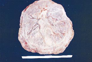

In a circumvallate (circummarginate) placenta the fetal surface may be reduced if decidual tissue has made its way between the amnion and chorion. This appears as a yellow, peripheral, hyalinized fold circumscribing the edge of the chorionic plate. This type of placenta has been reported to be a cause of antepartum bleeding and premature labor


This is an example of placenta membranacea (placenta diffusa). These placentas are rare. The ovum implants too deeply, the villae of the chorion fail to regress, and the placental tissue develops over the entire surface of the chorion. The placenta is very thin and is associated with poor fetal growth and antepartum hemorrhage. There may be previa type bleeding.

Thursday, June 29, 2017

Structural Abnormalities Of the Uterus

1. Arcuate uterus: 

  • The cavity has an indentation at the fundus.
  • This is probably the mildest form of uterine abnormality.


2. Septated Uterus: 

  • The uterus appears normal externally, but the cavity has a septum.

Thursday, May 18, 2017

Placental Abruption

 A 32 year old woman G4 P3+0 presents to the emergency department with heavy bleeding  and lower abdominal pain at 24 weeks gestation. On Examination there was no fetal heart beat, and the uterus was extremely tender on palpation. Ultrasound shows separation of the placenta, and a diagnosis of placental abruption was made which was managed accordingly. 


Placental Abruption - Discussion
In placental abruption also kniown as abruptio placentae , the placenta separates from the uterine wall prematurely, usually after the 20th week of gestation, producing hemorrhage. Abruptio placentae is most common in multigravidas—usually in women older than age 35—and is a common cause of bleeding during the second half of pregnancy. Fetal prognosis depends on gestational age and amount of blood lost; maternal prognosis is good if hemorrhage can be controlled.

Causes
In many cases, the cause of abruptio placentae is unknown. 

Predisposing factors include 
  • cocaine use, 
  • trauma (such as a direct blow to the uterus resulting from abuse or accidental trauma), 
  • placental site bleeding from a needle puncture during amniocentesis, 
  • chronic or pregnancy-induced hypertension (which raises pressure on the maternal side of the placenta), 
  • multiparity of more than five, 
  • short umbilical cord, 
  • dietary deficiency, 
  • smoking, 
  • advanced maternal age, and
  •  pressure on the venae cavae from an enlarged uterus.

Friday, September 30, 2016

A Case Of Pemphigoid Gestationis


A 37-year-old pregnant woman presented to the hospital with severe preeclampsia. After all medical methods were tried and failed to control her severe preeclampsia, a joint decision was made to induce
labor to save the life of the mother. The pregnancy was too early for the fetus to survive.
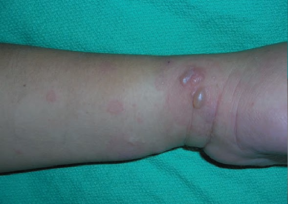
The following day she began to develop the lesions shown in picture above. Her past history includes antiphospholipid syndrome with multiple pregnancy losses. This was her third episode of having such skin lesions.
A diagnosis of pemphigoid gestationis was made after biopsy results from the lesions.

Case Discussion
 Pemphigoid Gestationis

Pemphigoid gestationis is a rare autoimmune bullous dermatosis of pregnancy. The disease was originally known as herpes gestationis because of it visual similarities to herpes simplex infection. However, that term has fallen out of favor because pemphigoid gestationis is not associated with active or prior herpes virus infection.

Pemphigoid gestationis, is defined as a bullous or blistering disease that is associated with pregnancy or with trophoblastic tumors

Pathology: 

• The pathophysiology of the disease involves immunoglobulin (Ig) G antibodies that attack cells in the skin.

Thursday, February 18, 2016

A 35 Year Old G5P4 Has A Retained Placenta After A Vaginal Delivery

A 35-year-old G5P4 woman at 39 weeks’ gestation is undergoing a vaginal delivery. She has a history of previous myomectomy and one prior low-transverse cesarean delivery. She was counseled about the risks, benefits, and alternatives of vaginal birth after cesarean, and elected a trial of labor. She proceeded through a normal labor. The delivery of the baby is uneventful. The placenta does not deliver after 30 minutes, and a manual extraction of the placenta is undertaken. The placenta seems to be firmly adherent to the uterus.

1. What is the most likely diagnosis?
2. What is your next step in management for this patient?


Answer And Discussion:

1. What is the most likely diagnosis?
Answer: Placenta accreta.

2. What is your next step in management for this patient?
Answer: Hysterectomy.

Case Discussion: 

Sunday, February 7, 2016

A Case Of Intra Uterine Growth Retardation

A 32-year-old primigravida is seen in your office at 33 weeks gestation for a routine prenatal visit. Her gestational age was calculated by her last normal menstrual period which was consistent with an ultrasound performed at 8 weeks gestation. Her pregnancy has been uneventful to date, although she has continued to smokeone pack or more of cigarettes daily. She states that she has been feeling normal fetal movement and no uterine contractions.
On examination, her height is 5 ft 6 in., her weight is 118 lb (53.5 kg), and her BP is 90/60 mm Hg. Her fundal height is 26 cm. On ultrasound, you note a single pregnancy with an estimated fetal weight of 900 g, which is at the 3rd percentile for gestational age.


  1. What is the most likely diagnosis?
  2. What other important items should be noted on the ultrasound?
  3.  What is the next step in the management of this patient?
  4. What are potential complications of the patient’s disorder?
Answers And Case Discussion: 

Tuesday, September 8, 2015

A Pregnant Woman Presents With Persistent Itching At 31 Weeks Of Gestation

A 32-year-old G3P2 woman presents with persistent itching in her 31st week of pregnancy. The itching is constant and worse at night. Her pregnancy had been uncomplicated and she has no past history of medical problems. Many excoriations are noted and there are no blister.

She has no jaundice or scleral icterus.  On laboratory workup her transaminases were greater than 300 and her total bilirubin was elevated at 2.1. Her bile salts were elevated and her hepatitis panel was negative. The ultrasound showed gallstones but no obstruction was seen.
A diagnosis of “intrahepatic cholestasis of pregnancy” was made and the patient was treated with oral ursodiol (a bile salt binding agent) and topical 1% hydrocortisone cream. The bile salts and transaminases were decreased and the patient’s pruritus improved but did not resolve until after delivery.

Case Discussion
Common Skin Findings In Pregnancy: 
Maternal skin and skin structures undergo numerous changes during pregnancy. There are two general categories of pregnancy-associated skin conditions:
(a) benign skin conditions associated with normal hormonal changes of pregnancy

  • Striae gravidarum, Or Stretch Marks
  • hyperpigmentation,
  • hair and vascular changes
(b) pregnancy-specific dermatoses

Sunday, February 1, 2015

21 Year Old female With Complaint Of Severe Right Lower Abdominal Pain

A 21 year old female presents to the emergency department with the complain of severe right lower abdominal pain that started this morning. The pain is 7/10 in intensity and constant in nature. It is exacerbated by movement and does not radiate. It is accompanied by fever, nausea and one episode of vomiting. Her LMP was 4 weeks back and she is sexually active. She has a history of regular periods every 4 weeks lasting for 6 days. She did notice some brownish spotting this morning which she thought was her periods because she was due. She is on birth control pills nowadays. She had a normal vaginal delivery 18 months back. Her past history is not significant for any illnesses or any major surgery.  On examination, she looked pale and was in pain. Her abdomen was tender on palpation more on the right side. On pelvic examination she had severe pain and the cervix and the uterus seemed to be pushed on one side.

 What will be the differential diagnosis on the basis of History and examination?

Differential Diagnosis:

  • Pelvic inflammatory disease.
  • Torsion of an ovary
  • Torsion of the ovarian cyst
  • Adnexal torsion
  • Acute appendicitis
  • Ruptured ectopic pregnancy
  • Abortion
 Urine hCG was negative and pregnancy was almost ruled out. She was taken for a transvaginal ultrasound and it shows the following picture;

Sonographic Whirlpool Sign in Ovarian Torsion


Thursday, January 15, 2015

A 23 Year Old Female With Oligomenorrhea

A 23 year old female presents to the clinic with presenting symptoms of  irregular menstrual periods ( every 3-4 months) and excessive facial hair. She is sexually active and never used birth control but has never been pregnant. On General appearance she looks obese and has acne.

The pelvic ultrasound is shown below:


The ultrasound shows multiple ovarian cysts. The patient is suffering from Polycystic ovarian Syndrome.

Case Discussion
Poly Cystic Ovarian Syndrome:

  • It is the most common cause of female hirsutism (male-pattern hair growth).
  • Patients may have mild or no symptoms or present with oligomenorrhea, infertilty and virilization.
  • The cause is umknown but hyperinsulinemia with insulin resistance is usually seen. 
  • Typically affects women in the teenage age years who are obese .