A 50-year-old man is seen in the Emergency department with severe central chest pain which he says is present for about 18 hours.
His ECG is shown below:
Question:
What does this ECG show and how would you manage this patient?
Answer:
Study and Memorize Medical Conditions With The Help Of Photos. Useful Site For Medical Students, Doctors And Nurses.
Showing posts with label Emergency Medicine. Show all posts
Saturday, June 24, 2017
Thursday, June 15, 2017
Traumatic Iridodialysis.
Traumatic iridodialysis is the result of an injury, typically blunt trauma that pulls the iris away from the ciliary body.
The resulting deformity appears as a lens-shaped defect at the outer margin of the iris.
Patients may present complaining of a “second pupil.” As the iris pulls away from the ciliary body,
a small amount of bleeding may result. Look closely for associated traumatic hyphema.
Consider etiologies such as
- penetrating injury to the globe,
- scleral rupture,
- Intraocular Foreign body and
- lens dislocation causing billowing of the iris.
Sunday, May 14, 2017
Fracture of Nose
The most common facial fracture, a fractured nose usually results from blunt injury and is commonly associated with other facial fractures. The severity of the fracture depends on the direction, force, and type of the blow.
A severe, comminuted fracture may cause extreme swelling or bleeding that may jeopardize the airway and require tracheotomy during early treatment. Inadequate or delayed treatment may cause permanent nasal displacement, septal deviation, and obstruction.
Causes
With low-energy injuries, noncomminuted nasal bone fragments are caused by low-velocity trauma. Such injuries could occur in the following situations:
Immediately after the injury, a nosebleed may occur, and soft-tissue swelling may quickly obscure the break. After several hours, pain, periorbital ecchymoses, and nasal displacement and deformity are prominent. A possible complication is septal hematoma, which may lead to abscess formation, resulting in vascular septic necrosis and saddle nose deformity.
A severe, comminuted fracture may cause extreme swelling or bleeding that may jeopardize the airway and require tracheotomy during early treatment. Inadequate or delayed treatment may cause permanent nasal displacement, septal deviation, and obstruction.
Causes
With low-energy injuries, noncomminuted nasal bone fragments are caused by low-velocity trauma. Such injuries could occur in the following situations:
- injuries created during fistfights (hand or fist blows only, no blunt instruments)
- uncomplicated falls such as tripping
- low-velocity motor vehicle collision.
- injuries sustained from a leveraged blow to the nose using an object such as a stick, pipe, or other blunt object
- falls from heights
- sport injuries with fast-moving projectiles, such as a ball or puck
- high-velocity motor vehicle collisions.
Immediately after the injury, a nosebleed may occur, and soft-tissue swelling may quickly obscure the break. After several hours, pain, periorbital ecchymoses, and nasal displacement and deformity are prominent. A possible complication is septal hematoma, which may lead to abscess formation, resulting in vascular septic necrosis and saddle nose deformity.
Monday, May 1, 2017
Acute Angle-Closure Glaucoma
Acute angle-closure glaucoma (ACG) is secondary to narrowing or closure of the anterior chamber angle, resulting in increased intraocular pressure (IOP), with subsequent damage to the optic nerve.
Pathophysiology: Normally, aqueous humor drains out of the anterior chamber via Schlemm canal in the anterior chamber angle. In ACG, this flow is impeded, and the IOP rises from a normal range of 10 to 21 mm Hg to 50 mm Hg or higher.
Clinical Presentation: ACG presents as an acutely inflamed eye. Nausea and vomiting are common and may be the presenting complaints. Eye pain and headache vary in severity. As the IOP reaches
the 50 to 60 mm Hg range, fluid is forced into the cornea, resulting in corneal edema. Patients report blurred vision and rainbow-colored halos around lights.
Acute Angle-Closure Glaucoma. The cornea is edematous, manifest by the indistinctness of the iris markings and the irregular corneal light reflex. Conjunctival hyperemia is also present.
Clinical findings include:
Pathophysiology: Normally, aqueous humor drains out of the anterior chamber via Schlemm canal in the anterior chamber angle. In ACG, this flow is impeded, and the IOP rises from a normal range of 10 to 21 mm Hg to 50 mm Hg or higher.
Clinical Presentation: ACG presents as an acutely inflamed eye. Nausea and vomiting are common and may be the presenting complaints. Eye pain and headache vary in severity. As the IOP reaches
the 50 to 60 mm Hg range, fluid is forced into the cornea, resulting in corneal edema. Patients report blurred vision and rainbow-colored halos around lights.
Acute Angle-Closure Glaucoma. The cornea is edematous, manifest by the indistinctness of the iris markings and the irregular corneal light reflex. Conjunctival hyperemia is also present.
Clinical findings include:
- tearing,
- perilimbal injection (“ciliary flush”),
- a cloudy (“steamy”) cornea,
- a nonreactive mid-dilated pupil,
- anterior chamber inflammation, and
- an increased IOP.
- Using a penlight or slit-lamp microscopy, the anterior chamber may appear shallow.
Monday, March 6, 2017
Uncal Herniation Syndrome After Severe Head Injuries
Severe head injury can result in extra-axial hematoma, cerebral contusion, or diffuse cerebral edema which, in turn, may cause one of five brain herniation syndromes:
Uncal herniation occurs when the uncus of the temporal lobe is displaced inferiorly through the medial edge of the tentorium.
Compression of cranial nerve III can cause an ipsilateral dilated pupil. Typically, patients with uncal herniation are unconscious and require intubation. A contusion to the eye may also result in a dilated, non responsive pupil and arouse suspicion for severe head injury and uncal herniation
but typically these patients will be alert.
Ipsilateral Dilated Pupil due to Uncal Herniation.
( CT revealed an epidural hematoma and unilateral effacement of the quadrigeminal cistern. )
Management: Intubate unconscious head trauma patients with a unilateral dilated pupil and transfer them immediately to a facility capable of caring for traumatic brain injury. A noncontrasted head CT scan can identify a subdural or epidural hematoma, diffuse edema, or temporal lobe contusion. These conditions often cause midline shift of cerebral structures and compression of the quadrigeminal cistern. Unilateral effacement of the quadrigeminal cistern confirms uncal herniation.
- uncal,
- central
- transtentorial,
- cerebellotonsillar,
- subfalcine, and external.
Uncal herniation occurs when the uncus of the temporal lobe is displaced inferiorly through the medial edge of the tentorium.
Compression of cranial nerve III can cause an ipsilateral dilated pupil. Typically, patients with uncal herniation are unconscious and require intubation. A contusion to the eye may also result in a dilated, non responsive pupil and arouse suspicion for severe head injury and uncal herniation
but typically these patients will be alert.
Ipsilateral Dilated Pupil due to Uncal Herniation.
( CT revealed an epidural hematoma and unilateral effacement of the quadrigeminal cistern. )
Management: Intubate unconscious head trauma patients with a unilateral dilated pupil and transfer them immediately to a facility capable of caring for traumatic brain injury. A noncontrasted head CT scan can identify a subdural or epidural hematoma, diffuse edema, or temporal lobe contusion. These conditions often cause midline shift of cerebral structures and compression of the quadrigeminal cistern. Unilateral effacement of the quadrigeminal cistern confirms uncal herniation.
Tuesday, February 14, 2017
Acute Right Ventricular Myocardial Infarction - ECG
ECG Findings
• ST elevation in right-sided V leads (V4R, V5R).
• ST elevation greater in lead III than lead II suggests RV MI.
• ST elevation in the normally obtained V1 also strongly suggests RV MI.
• Often associated with inferior MI and/or posterior MI.
ST elevation in V4R and V5R (arrows), with the V4 and V5 leads placed in their mirror-image locations on the right side of the chest. Any ST elevation seen in the right-sided precordial leads is significant.
Important Points:
1. The smaller muscle mass of the right ventricle produces a less intense injury pattern that is
External Ear Injuries - Brief Description With Pictures.
Injuries to the external ear may be open or closed.
- Blunt external ear trauma may cause a hematoma (otohematoma) of the pinna, which, if untreated, may result in cartilage necrosis and chronic scarring or further cartilage formation and permanent deformity (“cauliflower ear”).
- Open injuries include lacerations (with and without cartilage exposure) and avulsions
Pinna Hematoma. A hematoma has developed, characterized by swelling, discoloration, ecchymosis, and flocculence. Immediate incision and drainage or aspiration is indicated, followed by an ear compression dressing.
Management: Pinna hematomas must undergo incision and drainage or large needle aspiration using sterile technique, followed by a pressure dressing to prevent reaccumulation of the hematoma.
This procedure may need to be repeated several times; hence, after Emergency department drainage, the patient is treated with antistaphylococcal antibiotics and referred to ENT or plastic surgery for follow- up in 24 hours. Lacerations must be carefully examined for cartilage involvement; if this is present, copious irrigation, closure, and postrepair oral antibiotics covering skin flora are indicated.
Friday, February 10, 2017
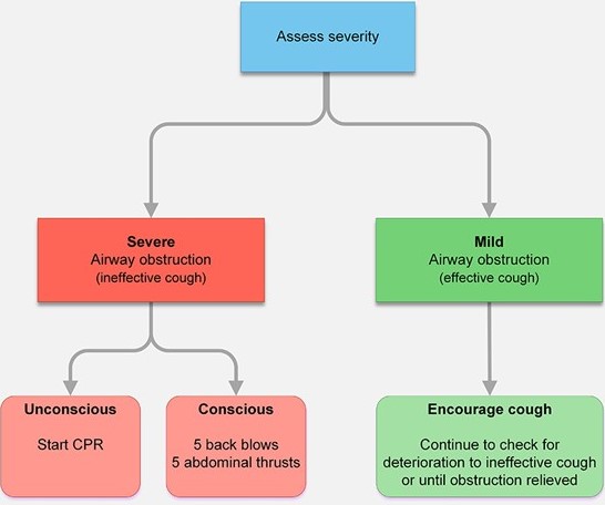
Choking In An Adult - Management Algorithm
The management of choking is rightly taught as part of first aid. Recognition of the problem is the key to success. Clues include a person experiencing a sudden airway problem whilst eating, possibly combined with them clutching their neck.Victims with severe airway obstruction may be unable to speak or breathe and become unconscious.
Monday, February 6, 2017
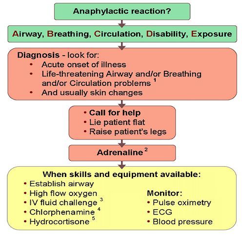
Treatment Algorithm And A Brief Description Of Anaphylaxis
Anaphylaxis is a generalized immunological condition of sudden onset, which develops after exposure to a foreign substance.
Pathophysiology: The mechanism may:
• Involve an IgE-mediated reaction to a foreign protein (stings, foods, streptokinase), or to a protein–hapten conjugate (antibiotics) to which the patient has previously been exposed.
• Be complement mediated (human proteins eg G -globulin, blood products).
• Be unknown (aspirin, ‘idiopathic’).
Irrespective of the mechanism, mast cells and basophils release mediators (eg histamine, prostaglandins, thromboxanes, platelet activating factors, leukotrienes) producing clinical manifestations.
Angio-oedema caused by ACE inhibitors and hereditary angio-oedema may present in a similar way
to anaphylaxis. Hereditary angio-oedema is not usually accompanied by urticaria and is treated with C1 esterase inhibitor.
Common causes: include:
Sunday, February 5, 2017
Depressed Skull Fracture
Depressed skull fractures typically occur when a large force is applied over a small area. They are classified as open if the skin above them is lacerated. Abrasions, contusions, and hematomas may also be present over the fracture site.
The patient’s mental status is dependent upon the degree of underlying brain injury. Direct trauma can cause abrasions, contusions, hematomas, and lacerations without an underlying depressed skull fracture. Evidence of other injuries such as a basilar fracture, facial fractures, or cervical spinal injuries may also be present.
CT demonstrating depressed skull fracture.
Management: Explore all scalp lacerations to exclude a depressed fracture. CT should be performed in all suspected depressed skull fractures to determine the extent of underlying brain injury.
The patient’s mental status is dependent upon the degree of underlying brain injury. Direct trauma can cause abrasions, contusions, hematomas, and lacerations without an underlying depressed skull fracture. Evidence of other injuries such as a basilar fracture, facial fractures, or cervical spinal injuries may also be present.
CT demonstrating depressed skull fracture.
Management: Explore all scalp lacerations to exclude a depressed fracture. CT should be performed in all suspected depressed skull fractures to determine the extent of underlying brain injury.
- Depressed skull fractures require immediate neurosurgical consultation.
- Treat open fractures with antibiotics and tetanus prophylaxis as indicated.
- The decision to observe or operate immediately is made by the neurosurgeon.
Saturday, February 4, 2017
Introduction To Erythema multiforme
Definition: Erythema multiforme (EM) is an acute, self-limited, and sometimes recurring skin condition that is considered to be a type IV hypersensitivity reaction.
Etiology: It occurs in response to medicines, infections, or illness
Clinical Features:
Erythema Multiforme. Symmetric distribution of targetoid macules and plaques. The dusky central zone is more obvious on the left waistline lesions.
Etiology: It occurs in response to medicines, infections, or illness
- Herpes simplex virus (HSV; frequently labialis) is strongly associated but may not be clinically apparent. Other viruses, bacteria (M pneumoniae, Chlamydia, Salmonella, Mycobacterium), and fungi (Histoplasma capsulatum, dermatophytes)
- are also associated.
- Medications account for <10%; NSAIDs, sulfonamides, antiepileptics, allopurinol, and antibiotics are
- responsible for the majority.
- Physical factors such as trauma, ultraviolet light exposure, and cold have been reported to elicit EM.
Clinical Features:
Erythema Multiforme. Symmetric distribution of targetoid macules and plaques. The dusky central zone is more obvious on the left waistline lesions.
- Erythema multiforme (EM) begins with symmetric, erythematous, sharply defined extremity or trunk macules, and evolves into a “targetoid” or “bull’s eye” morphology (a flat, dusky, central area with two concentric, erythematous rings).
- Bullae may appear in the central dusky area (bullous EM).
Shoulder Dislocation - A Brief DIscussion
Anterior shoulder dislocations are the most common and frequently caused by falling with the arm externally rotated and abducted.
Clinical Presentation: Patients present with the affected extremity held in adduction and internal rotation. Often, they complain of shoulder pain, refuse to move the affected arm, and may support the dislocated shoulder with the other arm. The acromion becomes prominent with loss of the rounded contour of the deltoid. A neurovascular examination of the upper extremity should be
performed to rule out associated injury, most commonly of the axillary nerve (sensation over the deltoid) and of the musculocutaneous nerve (sensation on the anterolateral forearm). Vascular injuries have rarely been reported to occur.
Diagnosis: Standard radiographic examination to evaluate for associated fracture should include AP and either axillary lateral or scapular views.
Anterior Shoulder Dislocation. Radiographic evaluation demonstrates that the humeral head is not in the glenoid fossa but is located anterior and inferior to it.
Posterior shoulder dislocations are commonly missed because of subtle radiographic findings.
Clinical Presentation: Patients present with the affected extremity held in adduction and internal rotation. Often, they complain of shoulder pain, refuse to move the affected arm, and may support the dislocated shoulder with the other arm. The acromion becomes prominent with loss of the rounded contour of the deltoid. A neurovascular examination of the upper extremity should be
performed to rule out associated injury, most commonly of the axillary nerve (sensation over the deltoid) and of the musculocutaneous nerve (sensation on the anterolateral forearm). Vascular injuries have rarely been reported to occur.
Diagnosis: Standard radiographic examination to evaluate for associated fracture should include AP and either axillary lateral or scapular views.
Anterior Shoulder Dislocation. Radiographic evaluation demonstrates that the humeral head is not in the glenoid fossa but is located anterior and inferior to it.
Posterior shoulder dislocations are commonly missed because of subtle radiographic findings.
Wednesday, February 1, 2017
Acromioclavicular Joint Seperation
Injury to the acromioclavicular (AC) joint usually results from an impact on the superior aspect of the acromion.
The classification system for AC joint injuries includes six types.
Clinical Features: Patients complain of pain at the AC joint and will actively splint the injured shoulder. Ecchymosis may be present; however, an obvious deformity is not always seen. There is significant tenderness upon palpation of the AC joint.
Diagnosis: Standard radiographs should include anteroposterior (AP) and axillary lateral views of the shoulder.
The classification system for AC joint injuries includes six types.
- A type I injury is equivalent to a stretching of the AC ligament.
- A type II injury consists of tearing of the AC ligaments and stretching of the coracoclavicular ligaments.
- Complete disruption of the AC and coracoclavicular ligaments is seen in types III to VI.
Clinical Features: Patients complain of pain at the AC joint and will actively splint the injured shoulder. Ecchymosis may be present; however, an obvious deformity is not always seen. There is significant tenderness upon palpation of the AC joint.
Diagnosis: Standard radiographs should include anteroposterior (AP) and axillary lateral views of the shoulder.
Testicular Torsion
A young male , age 16 years presents to the emergency with the complain of the sudden onset of pain in one testicle, followed by swelling of the affected testicle, reddening of the overlying scrotal skin, lower abdominal pain, nausea, and vomiting.
An examination reveals a swollen, tender, retracted testicle that often lies in the horizontal plane (bell-clapper deformity).
A diagnosis of Testicular torsion was made and the patient was prepared for immediate surgery.
Case Discussion:
Testicular torsion is one of the urologic emergencies and is very painful on presentation.
Testicular torsion occurs when a testicle rotates, twisting the spermatic cord that brings blood to the scrotum. The reduced blood flow causes sudden and often severe pain and swelling.
Testicular torsion is most common between ages 12 and 16, but it can occur at any age, even before birth.
An examination reveals a swollen, tender, retracted testicle that often lies in the horizontal plane (bell-clapper deformity).
A diagnosis of Testicular torsion was made and the patient was prepared for immediate surgery.
Case Discussion:
Testicular torsion is one of the urologic emergencies and is very painful on presentation.
Testicular torsion occurs when a testicle rotates, twisting the spermatic cord that brings blood to the scrotum. The reduced blood flow causes sudden and often severe pain and swelling.
Testicular torsion is most common between ages 12 and 16, but it can occur at any age, even before birth.
Tuesday, January 31, 2017
Tooth Subluxation
A 27 year old male was seen in the emergency department following a trauma and bleeding from the mouth. On examination he was found to have teeth subluxation due to the trauma.
Tooth Subluxation. Note the presence of blood along the crevice of the gingival margin of both central incisors—an indication of subluxation following trauma. Mild displacement of the subluxated teeth is noted
Case Discussion:
Tooth subluxation, the loosening of a tooth in its alveolar socket, is most commonly secondary to trauma; however, infection and periodontal disease may also produce subluxation.
Gingival lacerations and alveolar fractures are associated with dental subluxations. Gentle pressure to the teeth with a tongue blade or fingertip may produce movement, mild displacement, or blood along the crevice of the gingiva, all signs of subluxation.
Tooth Subluxation. Note the presence of blood along the crevice of the gingival margin of both central incisors—an indication of subluxation following trauma. Mild displacement of the subluxated teeth is noted
Case Discussion:
Tooth subluxation, the loosening of a tooth in its alveolar socket, is most commonly secondary to trauma; however, infection and periodontal disease may also produce subluxation.
Gingival lacerations and alveolar fractures are associated with dental subluxations. Gentle pressure to the teeth with a tongue blade or fingertip may produce movement, mild displacement, or blood along the crevice of the gingiva, all signs of subluxation.
Saturday, January 28, 2017
Basilar Skull Fracture
Introduction: The skull “base” comprises the frontal bone, occiput, occipital condyles, clivus, carotid canals, petrous portion of the temporal bones, and the posterior sphenoid wall. A basilar skull fracture is basically a linear fracture of the skull base.
Clinical Features: Trauma resulting in fractures to this area typically does not have localizing symptoms.
Indirect signs of the injury may include visible evidence of bleeding from the fracture into surrounding soft tissue, such as a Battle sign or “raccoon eyes.” (see picture shown below)
Battle Sign. A striking Battle sign is seen in this patient with head trauma. This finding may take hours to days to develop.
Bleeding into other structures—including hemotympanum or blood in the sphenoid sinus seen as an air-fluid level on computed tomography (CT)—may also be seen. Cerebrospinal fluid (CSF) leaks may also be evident and noted as clear or pink rhinorrhea. If CSF is present, a dextrose stick test may be positive. The fluid can be placed on filter paper and a “halo” or double ring may be seen.
Clinical Features: Trauma resulting in fractures to this area typically does not have localizing symptoms.
Indirect signs of the injury may include visible evidence of bleeding from the fracture into surrounding soft tissue, such as a Battle sign or “raccoon eyes.” (see picture shown below)
Battle Sign. A striking Battle sign is seen in this patient with head trauma. This finding may take hours to days to develop.
Bleeding into other structures—including hemotympanum or blood in the sphenoid sinus seen as an air-fluid level on computed tomography (CT)—may also be seen. Cerebrospinal fluid (CSF) leaks may also be evident and noted as clear or pink rhinorrhea. If CSF is present, a dextrose stick test may be positive. The fluid can be placed on filter paper and a “halo” or double ring may be seen.
Sunday, December 4, 2016
Toxic Epidermal Necrolysis
Toxic epidermal necrolysis (TEN) is a potentially lifethreatening skin disorder that is most commonly seen secondary to a drug reaction.
In this condition the skin develops a scalded appearance over an extensive area. Some authors consider TEN to be the severe end of a spectrum of skin disorders which includes erythema multiforme and Stevens Johnson syndrome
Clinical Features
- systemically unwell e.g. pyrexia, tachycardic
- positive Nikolsky's sign: the epidermis separates with mild lateral pressure
Drugs known to induce Toxic epidermal necrolysis (TEN)
Monday, November 21, 2016
Angioedema And Hereditary angioedema (HAE)
Regarding Angioedema answer the following questions:
1. What are the clinical pictures associated w ith angioedema?
2. What pathophysiologic processes underlie angioedema?
3. How is hereditary angioedema (HAE) diagnosed?
Answers:
1. What are the clinical pictures associated w ith angioedema?
2. What pathophysiologic processes underlie angioedema?
3. How is hereditary angioedema (HAE) diagnosed?
Answers:
Friday, November 11, 2016
Pleural Effusion- A Brief Discussion
CXR showing a large right pleural effusion
Pleural Effusion: Pleural effusion, sometimes referred to as “water on the lungs, is excess fluid that accumulates in the pleural cavity around the lungs.
The pleura are thin membranes that line the lungs and the inside of the chest cavity and act to lubricate and facilitate breathing. Normally, a small amount of fluid is present in the pleura.
Pleural Effusion: Pleural effusion, sometimes referred to as “water on the lungs, is excess fluid that accumulates in the pleural cavity around the lungs.
The pleura are thin membranes that line the lungs and the inside of the chest cavity and act to lubricate and facilitate breathing. Normally, a small amount of fluid is present in the pleura.
Causes: Numerous medical conditions can cause pleural effusions. Some of the more common causes are:
1.Transudative (watery fluid) pleural effusions:
- Heart failure
- Pulmonary embolism
- Cirrhosis
- Post open heart surgery
2. Exudative (protein-rich fluid) pleural effusions:
- Pneumonia
- Cancer
- Pulmonary embolism
- Kidney disease
- Inflammatory disease
Saturday, October 22, 2016
Subscribe to:
Posts (Atom)