Introduction:
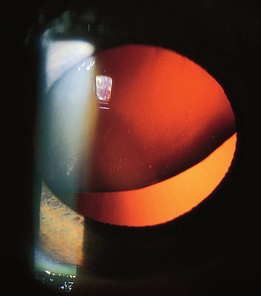
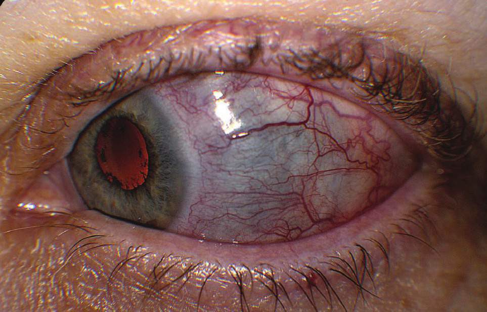
Any trauma to the eye that disrupts the normal architecture of the lens may result in the development of a traumatic cataract—a lens opacity.
The mechanism behind cataract formation involves fluid infiltration into the normally avascular and acellular lens stroma. The lens may be observed to swell with fluid and become cloudy and opacified. The time course is usually weeks to months following the original insult. Cataracts that are large enough may be observed by the naked eye. Those that are within the central visual field may cause blurring of vision or distortion of light around objects (eg, halos).
This acute traumatic cataract is seen as a milky cornea at the time of injury
Clinical Pearls:
1. Traumatic cataracts are frequent sequelae of lightning injury. Advise all victims of lightning strike of this possibility.
2. Cataracts may also occur as a result of electric current injury to the vicinity of the cranial vault.
3. Leukocoria results from a dense cataract, which causes loss of the red reflex.
4. If a cataract develops sufficient size and “swells” the lens, the trabecular meshwork may become blocked, producing glaucoma.
Study and Memorize Medical Conditions With The Help Of Photos. Useful Site For Medical Students, Doctors And Nurses.
Showing posts with label Ophthalmology. Show all posts
Friday, June 30, 2017
Friday, June 23, 2017
Lens Dislocation
Introduction
Lens dislocation may result from blunt trauma to the globe. As the anterior surface of the eye is struck, there is compression in the anteroposterior dimension with resultant stretching of the globe along its equator in the medial-lateral plane. As this occurs, it stretches the zonule fibers, which suspend the lens in place, and they along with the lens capsule may become disrupted.
Lens Dislocation. Lens dislocation revealed during slit-lamp examination. Note the zonule fibers, which normally hold the lens in place.
Clinical Features
The patient may experience symptoms of monocular diplopia or gross blurring of images, depending
on the severity of the injury. Occasionally there can be dramatic visual fluctuations caused by the lens changing position with resultant phakic and aphakic vision. There is generally a lack of pain except if secondary angle closure glaucoma occurs from the lens causing pupillary block.
Lens dislocation may result from blunt trauma to the globe. As the anterior surface of the eye is struck, there is compression in the anteroposterior dimension with resultant stretching of the globe along its equator in the medial-lateral plane. As this occurs, it stretches the zonule fibers, which suspend the lens in place, and they along with the lens capsule may become disrupted.
Lens Dislocation. Lens dislocation revealed during slit-lamp examination. Note the zonule fibers, which normally hold the lens in place.
Clinical Features
The patient may experience symptoms of monocular diplopia or gross blurring of images, depending
on the severity of the injury. Occasionally there can be dramatic visual fluctuations caused by the lens changing position with resultant phakic and aphakic vision. There is generally a lack of pain except if secondary angle closure glaucoma occurs from the lens causing pupillary block.
Thursday, June 15, 2017
Traumatic Iridodialysis.
Traumatic iridodialysis is the result of an injury, typically blunt trauma that pulls the iris away from the ciliary body.
The resulting deformity appears as a lens-shaped defect at the outer margin of the iris.
Patients may present complaining of a “second pupil.” As the iris pulls away from the ciliary body,
a small amount of bleeding may result. Look closely for associated traumatic hyphema.
Consider etiologies such as
- penetrating injury to the globe,
- scleral rupture,
- Intraocular Foreign body and
- lens dislocation causing billowing of the iris.
Saturday, June 10, 2017
Intraocular Foreign Body
The most important consideration with any eye injury is the possibility of a penetrating globe injury with residual intraocular foreign body (IO FB). Patients may report FB sensation, but subtle presentations occur. A meticulous history about the mechanism of injury (grinding or metal on metal) must be elicited.
Anterior Chamber Foreign Body. A shard from a nail is seen embedded in the anterior chamber. A “teardrop” pupil is present, indicating perforation
Clinical Pearls :
1. Always maintain a high index of suspicion for penetrating globe injury. Be particularly wary in mechanisms involving use of “metal on metal” such as grinding or hammering. A positive Seidel test demonstrates corneal microperforation.
2. If ocular penetration is suspected, a diligent search for a retained FB is indicated, beginning carefully with bedside ultrasound using a high-frequency transducer. Computed tomography (CT) is the diagnostic study of choice (avoid magnetic resonance imaging [MRI]) with indeterminate results or when confirmation is desired.
Anterior Chamber Foreign Body. A shard from a nail is seen embedded in the anterior chamber. A “teardrop” pupil is present, indicating perforation
Clinical Pearls :
1. Always maintain a high index of suspicion for penetrating globe injury. Be particularly wary in mechanisms involving use of “metal on metal” such as grinding or hammering. A positive Seidel test demonstrates corneal microperforation.
2. If ocular penetration is suspected, a diligent search for a retained FB is indicated, beginning carefully with bedside ultrasound using a high-frequency transducer. Computed tomography (CT) is the diagnostic study of choice (avoid magnetic resonance imaging [MRI]) with indeterminate results or when confirmation is desired.
Sunday, June 4, 2017
Hyphema - a pooling or collection of blood inside the anterior chamber of the eye .
Introduction
Injury to the anterior chamber that disrupts the vasculature supporting the iris or ciliary body results in a hyphema.
The blood tends to layer with time, and if left undisturbed, gravity will form a visible meniscus.
Definition: A hyphema is a pooling or collection of blood inside the anterior chamber of the eye (the space between the cornea and the iris).
Hyphema. This hyphema has completely layered out in the anterior chamber.
Clinical Symptoms can include
Injury to the anterior chamber that disrupts the vasculature supporting the iris or ciliary body results in a hyphema.
The blood tends to layer with time, and if left undisturbed, gravity will form a visible meniscus.
Definition: A hyphema is a pooling or collection of blood inside the anterior chamber of the eye (the space between the cornea and the iris).
Hyphema. This hyphema has completely layered out in the anterior chamber.
Clinical Symptoms can include
- pain,
- photophobia, and possibly blurred vision secondary to obstructing blood cells.
- Nausea and vomiting may signal a rise in intraocular pressure (glaucoma) caused by blood cells
- clogging the trabecular meshwork.
Monday, May 22, 2017
Description Of Exudates As Seen On Fundoscopy
Hard exudates are refractile, yellowish deposits with sharp margins composed of fat-laden macrophages and serum lipids.
Occasionally the lipid deposits form a partial or complete ring (called a circinate ring) around the leaking area of pathology. If the lipid leakage is located near the fovea, a spoke or star-type distribution of the hard exudates may be seen.
Hard Exudates. Linear collection of yellow lipid deposits with sharp margins in macula.
Cotton wool spots, or soft “exudates,” are actually microinfarctions of the retinal nerve fiber layer, and appear white with soft or fuzzy edges.
Cotton Wool Spots. White lesions with fuzzy margins, seen here approximately one-fifth to one-fourth disk diameter in size. Orientation of cotton wool spots generally follows the curvilinear
arrangement of the nerve fiber layer. Intraretinal hemorrhages and intraretinal vascular abnormalities are also present.
Inflammatory exudates are secondary to retinal or chorioretinal inflammation.
Occasionally the lipid deposits form a partial or complete ring (called a circinate ring) around the leaking area of pathology. If the lipid leakage is located near the fovea, a spoke or star-type distribution of the hard exudates may be seen.
Hard Exudates. Linear collection of yellow lipid deposits with sharp margins in macula.
Cotton wool spots, or soft “exudates,” are actually microinfarctions of the retinal nerve fiber layer, and appear white with soft or fuzzy edges.
Cotton Wool Spots. White lesions with fuzzy margins, seen here approximately one-fifth to one-fourth disk diameter in size. Orientation of cotton wool spots generally follows the curvilinear
arrangement of the nerve fiber layer. Intraretinal hemorrhages and intraretinal vascular abnormalities are also present.
Inflammatory exudates are secondary to retinal or chorioretinal inflammation.
Sunday, May 21, 2017
Sixth Nerve (Abducens Nerve) Palsy
The abducens nerve (Cranial Nerve VI) innervates the lateral rectus muscle and is the most common single muscle palsy, causing loss of abduction and resultant horizontal diplopia, worse in ipsilateral
gaze. Associated findings are dependent on the location of the lesion.
Etiology: Within the pons, involvement of the corticospinal tract results in contralateral hemiparesis. The abducens has the longest intracranial course of any nerve, and therefore is vulnerable to stretching or compression secondary to elevated intracranial pressure, trauma, neurosurgical manipulation, and cervical traction.
Also, any meningeal process (infectious, inflammatory, or neoplastic) can affect this portion of the sixth nerve.
Aneurysmal compression is uncommon.
Prior to entering the cavernous sinus, the nerve crosses the petrous portion of the temporal bone. Trauma with temporal bone fracture can result in a combination of sixth- and seventh-nerve palsies.
Cavernous sinus pathology is suggested by the involvement of the internal carotid artery, venous drainage of the eye and orbit, trochlear and oculomotor nerves, the first division of the trigeminal nerve, and the ocular sympathetics.
Microvascular changes secondary to diabetes, hypertension, and giant cell arteritis can compromise
function.
Sixth-Nerve Palsy. Loss of abduction of the left eye is seen in lateral gaze.
Management:
Associated signs and symptoms guide the workup.
gaze. Associated findings are dependent on the location of the lesion.
Etiology: Within the pons, involvement of the corticospinal tract results in contralateral hemiparesis. The abducens has the longest intracranial course of any nerve, and therefore is vulnerable to stretching or compression secondary to elevated intracranial pressure, trauma, neurosurgical manipulation, and cervical traction.
Also, any meningeal process (infectious, inflammatory, or neoplastic) can affect this portion of the sixth nerve.
Aneurysmal compression is uncommon.
Prior to entering the cavernous sinus, the nerve crosses the petrous portion of the temporal bone. Trauma with temporal bone fracture can result in a combination of sixth- and seventh-nerve palsies.
Cavernous sinus pathology is suggested by the involvement of the internal carotid artery, venous drainage of the eye and orbit, trochlear and oculomotor nerves, the first division of the trigeminal nerve, and the ocular sympathetics.
Microvascular changes secondary to diabetes, hypertension, and giant cell arteritis can compromise
function.
Management:
Associated signs and symptoms guide the workup.
Wednesday, May 17, 2017
Horner Syndrome - Clinical Features And Management
Horner syndrome (miosis, ptosis, and anhidrosis) is secondary to loss of ocular sympathetic innervation.
- Ptosis is less than 2 mm, the result of paralysis of Müller muscle, innervated by the sympathetic pathway.
- Anhidrosis is often not apparent to patients or clinicians.
- A pupillary finding specific in Horner syndrome is dilation lag. Because the dilator muscle is weak,
- the pupil dilates more slowly than the normal pupil.
This loss of ocular sympathetic innervation can be produced by a lesion anywhere along a three-neuron sympathetic pathway, from the hypothalamus down through the brain stem to the cervical cord, in the apex of the chest, along the carotid sheath, and in the cavernous sinus or orbit.
Isolated Horner syndrome presenting with head or neck pain suggests an internal carotid artery dissection.
Management
Associated signs and symptoms help direct the workup. A patient with cranial nerve abnormalities requires CT or MRI imaging and admission. In the setting of cervical spine trauma, neck immobilization and appropriate imaging studies are instituted. Consider carotid artery dissection in neck pain without trauma.
Sunday, May 14, 2017
Corneal Ulcer - Clinical features And Management
A corneal ulcer is an inflammatory and ulcerative keratitis.
Common infectious etiologies include
- Bacterial corneal ulcers are commonly associated with extended-wear contact lenses.
- Fungal infections may also arise from trauma involving vegetable matter such as a tree branch. - - - -Acanthamoeba infections may also occur from swimming in lakes, especially while wearing contact lenses.
Corneal Ulcer. A small circular corneal infiltrate is seen adjacent to the white flash photography reflection. Diffuse conjunctival hyperemia with a nasal ciliary flush is seen.
Clinical Features:Patients present with pain, photophobia, decreased vision, discharge, and a foreign body sensation.
Ocular findings include a corneal infiltrate, typically a round white spot, with conjunctival hyperemia, meiosis, and chemosis.
Slitlamp biomicroscopy may demonstrate an epithelial defect with fluorescein uptake. Anterior chamber findings can include cells and flare, keratic precipitates, and a hypopyon.
Common infectious etiologies include
- bacteria (Staphylococcus, Streptococcus, Pseudomonas) and
- viruses (herpes simplex, adenovirus).
- Rare causes of corneal ulcers include fungal infections and Acanthamoeba, a ubiquitous protozoan associated with contaminated contact lens solutions.
- Bacterial corneal ulcers are commonly associated with extended-wear contact lenses.
- Fungal infections may also arise from trauma involving vegetable matter such as a tree branch. - - - -Acanthamoeba infections may also occur from swimming in lakes, especially while wearing contact lenses.
Corneal Ulcer. A small circular corneal infiltrate is seen adjacent to the white flash photography reflection. Diffuse conjunctival hyperemia with a nasal ciliary flush is seen.
Clinical Features:Patients present with pain, photophobia, decreased vision, discharge, and a foreign body sensation.
Ocular findings include a corneal infiltrate, typically a round white spot, with conjunctival hyperemia, meiosis, and chemosis.
Slitlamp biomicroscopy may demonstrate an epithelial defect with fluorescein uptake. Anterior chamber findings can include cells and flare, keratic precipitates, and a hypopyon.
Wednesday, May 10, 2017
Ocular Herpes Simplex.
Ocular herpetic disease may be neonatal, primary, or recurrent.
Neonatal disease occurs secondary to passage through an infected birth canal, and is usually HSV type 2.
Primary ocular herpes may present as a blepharitis (grouped eyelid vesicles on an erythematous base), conjunctivitis, or keratoconjunctivitis.
Patients with keratoconjunctivitis commonly note pain, irritation, foreign body sensation, redness, photophobia, tearing, and occasionally decreased visual acuity. Follicles and preauricular adenopathy may be present. Initially, the keratitis is diffuse and punctate, but after 24 hours, fluorescein demonstrates either serpiginous ulcers or multiple diffuse epithelial defects. True dendritic ulcers are rarely seen in primary disease.
Ocular Herpes Simplex. This patient has a history of ocular herpes simplex since childhood. Grouped vesicles on an erythematous base with periorbital erythema are seen. Honey-colored crusts suggest secondary impetigo
Recurrent: Most ocular herpetic infections are manifestations of recurrent disease rather than a primary ocular infection.
These may be triggered by ultraviolet laser treatment, topical ocular medications (β-blockers, prostaglandins), and immunosuppression (especially ophthalmic topical glucocorticoids).
Recurrent disease most commonly presents as keratoconjunctivitis with a watery discharge, conjunctival injection, irritation, blurred vision, and preauricular lymph node involvement. Corneal involvement initially is punctate, but evolves into a dendritic keratitis. The linear branches classically end in bead-like extensions called terminal bulbs. Fluorescein dye demonstrates primarily the corneal defect; the terminal bulbs are best seen with rose stain. In addition to the dendritic pattern, fluorescein stain may instead take on a geographic or ameboid shape, secondary to widening of the
dendrite.
Neonatal disease occurs secondary to passage through an infected birth canal, and is usually HSV type 2.
Primary ocular herpes may present as a blepharitis (grouped eyelid vesicles on an erythematous base), conjunctivitis, or keratoconjunctivitis.
Patients with keratoconjunctivitis commonly note pain, irritation, foreign body sensation, redness, photophobia, tearing, and occasionally decreased visual acuity. Follicles and preauricular adenopathy may be present. Initially, the keratitis is diffuse and punctate, but after 24 hours, fluorescein demonstrates either serpiginous ulcers or multiple diffuse epithelial defects. True dendritic ulcers are rarely seen in primary disease.
Ocular Herpes Simplex. This patient has a history of ocular herpes simplex since childhood. Grouped vesicles on an erythematous base with periorbital erythema are seen. Honey-colored crusts suggest secondary impetigo
Recurrent: Most ocular herpetic infections are manifestations of recurrent disease rather than a primary ocular infection.
These may be triggered by ultraviolet laser treatment, topical ocular medications (β-blockers, prostaglandins), and immunosuppression (especially ophthalmic topical glucocorticoids).
Recurrent disease most commonly presents as keratoconjunctivitis with a watery discharge, conjunctival injection, irritation, blurred vision, and preauricular lymph node involvement. Corneal involvement initially is punctate, but evolves into a dendritic keratitis. The linear branches classically end in bead-like extensions called terminal bulbs. Fluorescein dye demonstrates primarily the corneal defect; the terminal bulbs are best seen with rose stain. In addition to the dendritic pattern, fluorescein stain may instead take on a geographic or ameboid shape, secondary to widening of the
dendrite.
Sunday, May 7, 2017
Herpes Zoster Opthalmicus
A middle aged lady presented with a vesicular rash on her face that was seen to be involving the ophthalmic division of the trigeminal nerve.
The case was diagnosed as Herpes Zoster Opthalmicus
Case Discussion:
Reactivation of endogenous latent varicella-zoster virus within the trigeminal ganglion with neuronal spread through the ophthalmic branch results in crops of grouped vesicles on the forehead and periocularly.
Clinical features; Patients typically present with periocular rash and an injected eye, along with a watery discharge. The most common corneal lesion is punctate epithelial keratitis, in which
the cornea has a ground-glass appearance because of stromal edema. Pseudodendrites, also very common, form from mucous deposition, are usually peripheral, and stain moderate to poorly with fluorescein. These may be differentiated from the dendrites of herpes simplex in that the pseudodendrites lack the rounded terminal bulbs at the end of the branches, and are broader and more plaquelike. Anterior stromal infiltrates may be seen in the second or third week after the acute
infection. Follicles (hyperplastic lymphoid tissue that appears as gray or white lobular elevations, particularly in the inferior cul-de-sac) and regional adenopathy may or may not be present. Iritis is seen in approximately 40% of patients.
The case was diagnosed as Herpes Zoster Opthalmicus
Case Discussion:
Reactivation of endogenous latent varicella-zoster virus within the trigeminal ganglion with neuronal spread through the ophthalmic branch results in crops of grouped vesicles on the forehead and periocularly.
Clinical features; Patients typically present with periocular rash and an injected eye, along with a watery discharge. The most common corneal lesion is punctate epithelial keratitis, in which
the cornea has a ground-glass appearance because of stromal edema. Pseudodendrites, also very common, form from mucous deposition, are usually peripheral, and stain moderate to poorly with fluorescein. These may be differentiated from the dendrites of herpes simplex in that the pseudodendrites lack the rounded terminal bulbs at the end of the branches, and are broader and more plaquelike. Anterior stromal infiltrates may be seen in the second or third week after the acute
infection. Follicles (hyperplastic lymphoid tissue that appears as gray or white lobular elevations, particularly in the inferior cul-de-sac) and regional adenopathy may or may not be present. Iritis is seen in approximately 40% of patients.
Saturday, May 6, 2017
Anisocoria
Anisocoria is a disparity of pupil size.
To determine the abnormal pupil, compare pupil sizes in light and dark. It is accentuated in the paretic muscle. If the iris sphincter (or its innervation) is involved, the anisocoria will be increased in bright light. If the iris dilator muscle (or its innervation) is affected, it will be more
pronounced in darkness.
Up to 20% of normal individuals have physiologic anisocoria of 1 to 2 mm.
Other causes of anisocoria with an abnormally large pupil include
- mydriatic drops,
- contamination from a scopolamine patch,
- an Adie pupil, and
- ocular trauma with iris sphincter damage.
Post traumatic Anisocoria. Marked chronic anisocoria secondary to prior trauma as a child
A dilated pupil due to anticholinergic agents (eg, atropine) does not react to light, although a dilated pupil due to sympathomimetics still has some response.
An abnormally small pupil may be secondary to Horner syndrome, chronic Adie pupil (8 weeks or more after the event), iritis, and eye drops (pilocarpine).
Miosis secondary to pupillary sphincter muscle spasm may be transiently observed after ocular trauma, followed by mydriasis.
To determine the abnormal pupil, compare pupil sizes in light and dark. It is accentuated in the paretic muscle. If the iris sphincter (or its innervation) is involved, the anisocoria will be increased in bright light. If the iris dilator muscle (or its innervation) is affected, it will be more
pronounced in darkness.
Up to 20% of normal individuals have physiologic anisocoria of 1 to 2 mm.
Other causes of anisocoria with an abnormally large pupil include
- mydriatic drops,
- contamination from a scopolamine patch,
- an Adie pupil, and
- ocular trauma with iris sphincter damage.
Post traumatic Anisocoria. Marked chronic anisocoria secondary to prior trauma as a child
A dilated pupil due to anticholinergic agents (eg, atropine) does not react to light, although a dilated pupil due to sympathomimetics still has some response.
An abnormally small pupil may be secondary to Horner syndrome, chronic Adie pupil (8 weeks or more after the event), iritis, and eye drops (pilocarpine).
Miosis secondary to pupillary sphincter muscle spasm may be transiently observed after ocular trauma, followed by mydriasis.
Monday, May 1, 2017
Anterior Uveitis (Iritis)
Introduction: The uvea is the middle layer of the eye. Its anterior portion includes the iris and ciliary body; the posterior portion includes the choroid. Inflammation of the anterior portion is called anterior uveitis or iritis.
Etiology: It is often idiopathic, but approximately half of cases are associated with systemic
disease. These include :
Clinical features include conjunctival hyperemia, hyperemic perilimbal vessels (“ciliary flush”), miosis, decreased visual acuity, photophobia, tearing, and pain.
Anterior Uveitis. Marked conjunctival injection and perilimbal hyperemia (“ciliary flush”) are seen in this patient with recurrent iritis
The slit-lamp may demonstrate a hypopyon, cells, flare, and keratic precipitates.
Keratic precipitates are agglutinated inflammatory cells adherent to the posterior corneal endothelium. These precipitates appear either as fine gray-white deposits or as a large, flat, greasy-looking area (“mutton fat”).
The IOP may be decreased due to decreased aqueous production by the inflamed ciliary body, or increased secondary to inflammatory debris within the trabeculae of the anterior chamber angle obstructing outflow.
Etiology: It is often idiopathic, but approximately half of cases are associated with systemic
disease. These include :
- Inflammatory disorders (rheumatoid arthritis, Behçet disease, sarcoid),
- HLA-B27-associated conditions (ankylosing spondylitis, inflammatory bowel disease, Reiter syndrome), and
- Infectious causes (zoster, tuberculosis, toxoplasmosis, AIDS).
Clinical features include conjunctival hyperemia, hyperemic perilimbal vessels (“ciliary flush”), miosis, decreased visual acuity, photophobia, tearing, and pain.
Anterior Uveitis. Marked conjunctival injection and perilimbal hyperemia (“ciliary flush”) are seen in this patient with recurrent iritis
The slit-lamp may demonstrate a hypopyon, cells, flare, and keratic precipitates.
Keratic precipitates are agglutinated inflammatory cells adherent to the posterior corneal endothelium. These precipitates appear either as fine gray-white deposits or as a large, flat, greasy-looking area (“mutton fat”).
The IOP may be decreased due to decreased aqueous production by the inflamed ciliary body, or increased secondary to inflammatory debris within the trabeculae of the anterior chamber angle obstructing outflow.
Acute Angle-Closure Glaucoma
Acute angle-closure glaucoma (ACG) is secondary to narrowing or closure of the anterior chamber angle, resulting in increased intraocular pressure (IOP), with subsequent damage to the optic nerve.
Pathophysiology: Normally, aqueous humor drains out of the anterior chamber via Schlemm canal in the anterior chamber angle. In ACG, this flow is impeded, and the IOP rises from a normal range of 10 to 21 mm Hg to 50 mm Hg or higher.
Clinical Presentation: ACG presents as an acutely inflamed eye. Nausea and vomiting are common and may be the presenting complaints. Eye pain and headache vary in severity. As the IOP reaches
the 50 to 60 mm Hg range, fluid is forced into the cornea, resulting in corneal edema. Patients report blurred vision and rainbow-colored halos around lights.
Acute Angle-Closure Glaucoma. The cornea is edematous, manifest by the indistinctness of the iris markings and the irregular corneal light reflex. Conjunctival hyperemia is also present.
Clinical findings include:
Pathophysiology: Normally, aqueous humor drains out of the anterior chamber via Schlemm canal in the anterior chamber angle. In ACG, this flow is impeded, and the IOP rises from a normal range of 10 to 21 mm Hg to 50 mm Hg or higher.
Clinical Presentation: ACG presents as an acutely inflamed eye. Nausea and vomiting are common and may be the presenting complaints. Eye pain and headache vary in severity. As the IOP reaches
the 50 to 60 mm Hg range, fluid is forced into the cornea, resulting in corneal edema. Patients report blurred vision and rainbow-colored halos around lights.
Acute Angle-Closure Glaucoma. The cornea is edematous, manifest by the indistinctness of the iris markings and the irregular corneal light reflex. Conjunctival hyperemia is also present.
Clinical findings include:
- tearing,
- perilimbal injection (“ciliary flush”),
- a cloudy (“steamy”) cornea,
- a nonreactive mid-dilated pupil,
- anterior chamber inflammation, and
- an increased IOP.
- Using a penlight or slit-lamp microscopy, the anterior chamber may appear shallow.
Thursday, April 27, 2017
Episcleritis - A Brief Introduction
Introduction: Episcleritis is a common and benign inflammation of the episclera, typically affecting young and middle-aged adults. Seventy percent of cases occur in females.
The episclera lies just beneath the bulbar conjunctiva. Its vessels are large, run in a radial direction,
and can be seen beneath the overlying conjunctiva. Episcleral and conjunctival vessels blanch with the use of topical 5% phenylephrine drops, unlike deep episcleral vessels.
Clinical Features:
- Patients may complain of foreign body sensation, mild tenderness, irritation, mild photophobia, and excessive lacrimation.
- Pain is unusual but can occur, particularly in chroniccases.
- One-half of cases are bilateral.
- Eye findings are notable for a localized pink or bright red conjunctival injection, with involvement of the vessels in the superficial episcleral vascular plexus.
- Visual acuity is normal.
Associated Conditions: Episcleritis is usually an isolated condition, though it may be associated with a number of systemic diseases, including rheumatoid arthritis, inflammatory bowel disease, lupus, and vasculitis.
Scleritis
Definition: Scleritis is defined as inflammation of the sclera ( which is the white outer coating of the eye).Scleritis is painful, destructive, and potentially blinding condition of the eye.
Clinical features: The pain is constant and boring and may radiate to the face and periorbital
region. Associated features include tearing, photophobia, globe tenderness to palpation, and painful ocular movement.
The conjunctival vessels are injected. The eye itself may be intensely red with a violaceous or purple hue secondary to engorgement of the deep vessels of the episclera and scleral thinning. These deep vessels do not move when the overlying conjunctiva is moved with a cotton-tipped applicator, nor do
they blanch with topical phenylephrine.
On slit-lamp microscopy, the episcleral vessels are displaced outward by scleral edema.
Corneal involvement, iritis (with cells and flare in the anterior chamber), and decreased visual acuity may accompany scleritis.
Scleritis. A 55-year-old female with scleritis of the left eye associated with rheumatoid arthritis. Note dilation of the deep conjunctival and episcleral vessels and blue hue suggesting thinning of the sclera temporally
Etiology: An associated scleritis may occur with severe infectious keratitis. Primary infectious
Clinical features: The pain is constant and boring and may radiate to the face and periorbital
region. Associated features include tearing, photophobia, globe tenderness to palpation, and painful ocular movement.
The conjunctival vessels are injected. The eye itself may be intensely red with a violaceous or purple hue secondary to engorgement of the deep vessels of the episclera and scleral thinning. These deep vessels do not move when the overlying conjunctiva is moved with a cotton-tipped applicator, nor do
they blanch with topical phenylephrine.
On slit-lamp microscopy, the episcleral vessels are displaced outward by scleral edema.
Corneal involvement, iritis (with cells and flare in the anterior chamber), and decreased visual acuity may accompany scleritis.
Scleritis. A 55-year-old female with scleritis of the left eye associated with rheumatoid arthritis. Note dilation of the deep conjunctival and episcleral vessels and blue hue suggesting thinning of the sclera temporally
Etiology: An associated scleritis may occur with severe infectious keratitis. Primary infectious
Wednesday, April 26, 2017
Dacryoadenitis - Case Discussion
This young patient is referred to the ophthalmologist with the presenting complains of unilateral upper eyelid swelling and tenderness.
The case of diagnosed as Dacryoadenitis.
Case Discussion :
Dacryoadenitis is the inflammation of the lacrimal glands
Acute dacryoadenitis typically involves children and young adults with associated systemic infections such as gonorrhea, mumps, Epstein-Barr virus, and Staphlyococcus species.
Clinical Findings are localized to the outer one-third of the upper eyelid and include fullness or swelling, erythema, and tenderness.
A characteristic “S”-shaped deformity with ptosis of the lid may be seen.
In more advanced cases proptosis, inferonasal globe displacement, ophthalmoparesis, and diplopia may be present.
Chronic dacryoadenitis is more common, is seen in older patients, and is usually due to tumor or associated inflammatory disorders such as sarcoidosis, Sjögren syndrome, or IgG4-related diseases.
Dacryocystitis - Clinical Features And Management.
Definition: Dacryocystitis is an inflammation of the lacrimal sac, positioned immediately distal to the canaliculi and proximal to the nasolacrimal duct.
Etiology: Inflammation is usually secondary to obstruction of the nasolacrimal duct.
Clinical findings include:
During examination tears and mucopurulence may be expressed from the punctum when pressure is applied over the lacrimal sac.
Dacryocystitis. Swelling and erythema over the medial lower lid and lacrimal sac developed in this 10-year-old patient with streptococcal pharyngitis.
Complications include:
Up to 20% of normal newborns have a closed nasolacrimal passage, and 90% spontaneously open within the first 6 months.
Etiology: Inflammation is usually secondary to obstruction of the nasolacrimal duct.
Clinical findings include:
- swelling over the lacrimal sac,
- redness,
- tearing,
- eyelash matting and crusting,
- conjunctival redness.
During examination tears and mucopurulence may be expressed from the punctum when pressure is applied over the lacrimal sac.
Dacryocystitis. Swelling and erythema over the medial lower lid and lacrimal sac developed in this 10-year-old patient with streptococcal pharyngitis.
Complications include:
- conjunctivitis and
- orbital or preseptal cellulitis.
Up to 20% of normal newborns have a closed nasolacrimal passage, and 90% spontaneously open within the first 6 months.
Monday, April 24, 2017
Allergic Conjunctivitis
Allergic conjunctivitis is a condition whereby airborne allergens precipitate type 1 IgE-mediated hypersensitivity reactions in the conjunctiva.
Allergens include:
- pollens,
- animal dander,
- mites,
- mold, and
- dust.
Approximately 50% of patients have a personal or family history of allergic conditions such as
atopy, eczema, asthma, and allergic rhinitis.
Clinical Features: Itching is the hallmark symptom. Associated clinical features include:
- conjunctival injection and edema,
- burning,
- discharge (clear, white, or mucopurulent),
- chemosis, and
- eyelid redness and swelling.
- Small papillae may be seen on the tarsal conjunctiva.
Vernal conjunctivitis is a rare but serious form of allergic conjunctivitis. The highest incidence is seen in the arid areas of the Middle East and North Africa secondary to wind and dust storms. Symptoms are similar to allergic conjunctivitis, but are more intense. Itching is severe and a vigorous knuckle rubbing is a typical observation. Giant, raised, pleomorphic papillae (“cobblestones”)
seen over the upper tarsal plate are pathognomonic.
Tuesday, April 18, 2017
A Brief Discussion On Viral Conjunctivitis
Viral Conjunctivitis. Note the characteristic asymmetric conjunctival injection. Symptoms first developed in the left eye, with symptoms spreading to the other eye a few days later. A thin watery discharge is also seen.
Viral conjunctivitis is a common presentation of the red eye.
Findings are mild and include :
The tarsal conjunctiva may appear bumpy secondary to hyperplastic lymphoid tissue (follicles). Preauricular adenopathy may be present. The visual acuity is normal. The infection usually begins in one eye, but both eyes usually become involved due to autoinoculation.
There are few to no systemic complaints.
Adenovirus is the most common virus. A point of care test now available may aid clinicians to avoid empiric antibiotic therapy.
Epidemic keratoconjunctivitis (EKC) is a severe and highly contagious adenovirus infection that also involves the cornea. Additional features may include:
Viral conjunctivitis is a common presentation of the red eye.
Findings are mild and include :
- a thin watery discharge,
- crusting in the morning,
- burning or irritation,
- conjunctival injection (typically diffuse), and
- lid edema.
The tarsal conjunctiva may appear bumpy secondary to hyperplastic lymphoid tissue (follicles). Preauricular adenopathy may be present. The visual acuity is normal. The infection usually begins in one eye, but both eyes usually become involved due to autoinoculation.
There are few to no systemic complaints.
Adenovirus is the most common virus. A point of care test now available may aid clinicians to avoid empiric antibiotic therapy.
Epidemic keratoconjunctivitis (EKC) is a severe and highly contagious adenovirus infection that also involves the cornea. Additional features may include:
Subscribe to:
Posts (Atom)