Showing posts with label Pediatrics. Show all posts

Tuesday, September 18, 2018

The Placenta, Its Membranes, and the Umbilical Cord

The human placenta is a highly sophisticated organ of interface between mother and fetus, often
referred to as the “gate-keeper to the fetus.” Careful examination of the placenta, its membranes,
and the umbilical cord can prove to be a valuable aid in the diagnosis and treatment of the
neonate. Gross examination of the placenta takes five minutes, and more sophisticated examination
should be considered when there is poor pregnancy outcome, recognizable malformations or
abnormalities, multiple gestation, extremes of amniotic fluid volume, severe intrauterine growth
retardation, short umbilical cord (< 32 cm), and profound acidemia.
The maternal surface of the placenta (decidual plate) is soft, spongy and dark red; and the fetal surface (chorionic plate) is shiny and steel blue to gray.
The placenta, membranes, and umbilical cord weigh approximately 400 to 600 g at birth.

Abnormalities in structure can result in an inefficient transport of oxygen and nutrients to the developing baby. Despite this importance, it is one of the least understood and investigated human organs.

A succenturiate (accessory) lobe is common and has no effect on the fetus. This occurs in about 3 to 5% of deliveries.  Its importance arises from the fact that it may be retained within the uterus and cause postpartum bleeding.


Fetal surface of a bipartite or bilobed placenta (placenta duplex). The two parts of the placenta
are of nearly equal size and this occurs in about 1% of deliveries. Note that the lobes are separated by membranes. The umbilical cord may insert into one or other lobe, or may insert between the two.
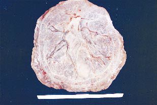

In a circumvallate (circummarginate) placenta the fetal surface may be reduced if decidual tissue has made its way between the amnion and chorion. This appears as a yellow, peripheral, hyalinized fold circumscribing the edge of the chorionic plate. This type of placenta has been reported to be a cause of antepartum bleeding and premature labor


This is an example of placenta membranacea (placenta diffusa). These placentas are rare. The ovum implants too deeply, the villae of the chorion fail to regress, and the placental tissue develops over the entire surface of the chorion. The placenta is very thin and is associated with poor fetal growth and antepartum hemorrhage. There may be previa type bleeding.

Monday, October 30, 2017

Some Limb Deformities in a Newborn

1. Metatarsus adductus. In metatarsus adductus, the forefoot is deviated medially and is slightly supinated. In the normal foot, a line drawn through the hindfoot will pass between the second and third toe. With metatarsus adductus, this line will pass lateral to the third toe. The prognosis is excellent, with most cases of metatarsus adductus resolving spontaneously.


2. Calcaneovalgus. Calcaneovalgus deformity of the foot is commonly associated with lateral tibial torsion. The forefoot is abducted, and the ankle is severely dorsiflexed to where the foot folds against the anterolateral surface of the tibia. The deformity is usually flexible, and the foot can be passively placed in the normal position. Calcaneovalgus deformity of the foot usually resolves spontaneously.


3. Talipes equinovarus. Talipes equinovarus, or club foot, has an incidence of approximately 1.5 in 1,000. Fifty percent of the time, the condition is bilateral. It can be associated with other conditions such as spina bifida and arthrogryposis. The foot turns inward and downward and remains tight in this position. Talipes equinovarus requires immediate evaluation since the timing of corrective casting, if necessary, can affect optimal outcome.

Friday, June 30, 2017

Meconium Aspiration Syndrome - X ray Chest

This X ray is of a one day old baby who had difficulty breathing.

X-ray chest shows right upper lobe consolidation and rest of the lungs show patchy to streaky areas suggesting meconium aspiration syndrome

Explanation: In meconium aspiration syndrome (MAS) the newborn breathes a mixture of meconium and amniotic fluid into the lungs around the time of delivery.
Meconium is the early feces passed by a newborn soon after birth, in some cases; the baby passes meconium while still inside the uterus. This usually happens due to lack of enough blood and oxygen.
Once the meconium has passed into the amniotic fluid, the baby may breathe meconium into the lungs. This condition is called MAS. It causes breathing difficulty due to inflammation in the lungs after birth.
Meconium aspiration syndrome is a leading cause of severe illness and death in newborns. However, in most cases, the outlook is excellent and there are no long-term health effects provided the infant is placed in newborn intensive care unit for close observation and treatment

Monday, June 19, 2017

Neonatal Dermatoses - Different Types Explained with Pictures For Better Understanding.

Skin disorders are one of the most common problems in pediatrics. Never underestimate parental concerns about their child’s skin. Unlike many disease processes, the skin is visible and noticeable to parents and others. Examination of the skin requires observation and palpation of the entire skin surface under good light. Do not forget to look at the eyes and mouth for mucous membrane involvement. Examination should include onset, duration, and inspection of a primary lesion. It is also important to note secondary changes, morphology, and distribution of the lesions.

Neonatal Dermatoses

1. Cutis Marmorata

  • Transient, blanchable, reticulated mottling occurs on the skin exposed to a cool environment.
  • No treatment is necessary; the condition generally resolves by 1 year of age.
  • If it persists, consider hypothyroidism, heart disease, or other associated abnormalities.
2. Erythema Toxicum Neonatorum

  • Scattered erythematous papules and pustules may occur anywhere on the body .
  • This self-limited condition generally appears in the first week of life and resolves within 1 month.

Thursday, June 15, 2017

A 2 year old child with Bronchopneumonia

A 2 years old child was brought to radiology department for X-ray chest with cough and high fever over last 3 days. His X ray is shown below:


X-ray chest shows soft small ill-defined opacities scattered in both lung fields partially sparing the lower zones.

Comments And Explanation
On X-ray bronchopneumonia appears as small fluffy ill-defined acinar nodules, which coalesce and enlarge with time and may develop into segmental and lobar densities with volume loss from airway obstruction secondary to mucus plugging and bronchial narrowing.

Diagnosis: Bronchopneumonia.

Clinical Discussion
Bronchopneumonia is a combination of interstitial and alveolar disease.
In bronchopneumonia the insult begins in airways, involves bronchovascular bundle and trickles into alveoli, which may develop and contain edema fluid, blood, leukocytes, hyaline membranes and organisms.

Thursday, May 18, 2017

A Newborn Baby with Difficulty Breathing since Birth

X ray of a newborn baby with difficulty breathing since birth is shown below:

Radiological Findings:  X-ray chest  shows opaque left hemithorax and the entire mediastinum is pulled to the left, the right lung has herniated beyond the midline. The endotracheal tube and Ryles tube are in situ. 

Diagnosis suggested is complete agenesis of left lung. This can be confirmed on CT chest.

Comments: 
The findings seen on chest film are absence of lung, opaque hemithorax, mediastinal shift with hernia of contralateral lung to the affected side with normal and symmetrical bony thorax.

CT chest can be helpful to differentiate the three categories of agenesis
(a) complete agenesis, 
(b) lung aplasia and 
(c) lung hypoplasia.
Lung agenesis is rare, developmental anomaly of the lung. It can be:
1. Complete agenesis, manifested by total agenesis of lung tissue, bronchus and pulmonary vessels.
2. Lung aplasia manifested by a rudimentary bronchus but absent lung tissue and pulmonary vessels.
3. Lung hypoplasia manifested by rudimentary bronchus with hypoplastic lung tissue and pulmonary vessels.

Sunday, May 7, 2017

Introduction to Mumps


Also known as infectious or epidemic parotitis, mumps is an acute viral disease caused by a paramyxovirus. It’s most prevalent in unvaccinated children between ages 2 and 12, but it can occur in other age-groups. Infants younger than age 1 seldom get this disease because of passive immunity from maternal antibodies. Peak incidence occurs during late winter and early spring.

The prognosis for complete recovery is good, although mumps sometimes causes complications.

Etiology
The mumps paramyxovirus is found in the saliva of an infected person and is transmitted by droplets or by direct contact. The virus is present in the saliva 6 days before to 9 days after onset of parotid gland swelling; the 48-hour period immediately preceding onset of swelling is probably the time of highest communicability.

The incubation period ranges from 14 to 25 days (the average is 18 days). One attack of mumps (even if unilateral) almost always confers lifelong immunity.

Signs and symptoms
Signs and symptoms of mumps vary widely. An estimated 30% of susceptible people have subclinical illness. Mumps usually begins with prodromal signs and symptoms that last for 24 hours; these include myalgia, anorexia, malaise, headache, and low-grade fever, followed by an earache that’s aggravated by chewing, parotid gland tenderness and swelling, a temperature of 101° to 104° F (38.3° to 40° C), and pain when chewing or when drinking sour or acidic liquids. Simultaneously with the swelling of the parotid gland or several days later, one or more of the other salivary glands may become swollen.

Tuesday, February 7, 2017

Nevus Simplex Or A Salmon Patch - A Vascular Lesion Of Infancy

Nevus simplex (salmon patch) is the most common vascular lesion in infancy, present in about 40% to 60% of newborns. It appears as a blanching, slightly pink-red macule or patch most commonly on the nape of the neck, the glabella, mid-forehead, or upper eyelids. Lesions generally fade over the first 2 years of life but may become more prominent with crying or straining.

                                 Newborn with characteristic salmon patches over his face.

Important Points:
1. Salmon patches are composed of ectatic dermal capillaries.
2. Salmon patches appear symmetrically and cross the midline in contrast to the unilateral distribution of a port-wine stain.
3. When seen on the nape of the neck, this lesion is referred to as a stork bite  (see picture below) or as an angel’s kiss when appearing on the forehead.

Wednesday, February 1, 2017

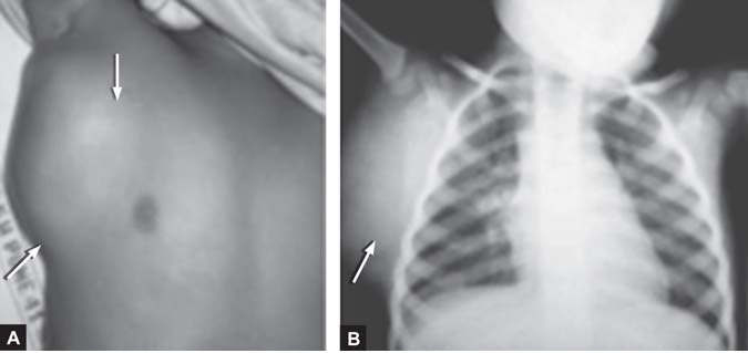
A 3 Year Old Child With A Mass On The Chest

A 3 years old child was seen by the pediatrician for a large soft tissue mass on right side of chest.

Photograph (A) of the child shows the soft mass (arrow), on X-ray chest (B) the soft tissue lymph nodal mass lies outside the thoracic cage in right hemithorax.

Diagnosis: Lymph node mass.

Discussion: On X-ray chest the lung fields are clear, on X-ray chest it is important to look for any lesion in the soft tissue over the entire available film. It may be in the form of enlarged thyroid gland, cervical or axillary lymph nodes, neurofibroma, surgical emphysema, a lesion in the breast or one
of the breasts might have been surgically excised. Ultrasound can be useful in investigating nodal masses particularly as an aid to biopsy.

Sunday, January 29, 2017

Neonatal Conjunctivitis - A Brief Discussion

Neonatal conjunctivitis is acquired either during birth with passage through the mother’s cervix and vagina, or from cross-infection in the neonatal period.

Etiologies: More common etiologies include Chlamydia trachomatis, viruses (herpes simplex), and bacteria (Staphylococcus aureus, Streptococcal pneumoniae, Haemophilus species).

Clinical Features: 

  • Presenting symptoms for Neisseria gonorrhoeae infection include a hyperacute bilateral conjunctivitis with copious purulent discharge, lid swelling, chemosis, and preauricular adenopathy.

            Thick purulent drainage in a newborn diagnosed with neonatal gonococcal conjunctivitis.

  • For chlamydial conjunctivitis, the clinical features range from mild swelling with a watery discharge to marked lid swelling with a red and thickened conjunctiva with a blood-stained discharge. 
  • Fluorescein staining of herpes simplex conjunctivitis demonstrates epithelial dendrites.
  • Blindness can result from gonococcal eye infection in the neonate because the organism can invade the cornea. It is one of the few emergency conjunctival infections.
  •  Nasolacrimal duct obstruction is common (up to 20%) in newborns and may present with findings suggestive of conjunctivitis. It is a diagnosis of exclusion in the neonate.

Tuesday, November 29, 2016

A Child Presents With A Skin Lesion With No Other Associated Symptoms.

A 6-year-old child presents with the skin lesions shown below:


The lesions have been present over the last month, and the child has reported no symptoms associated with them. The most likely diagnosis is
A) Varicella
B) Herpes zoster
C) Rhus dermatitis
D) Molluscum contagiosum
E) Scabies

The answer is