Showing posts with label MRCP Practice Questions. Show all posts

Wednesday, February 15, 2017

A 19 Year Old Woman With Fever And Sore Throat

A 19-yr-old woman presents with a severe sore throat, fever and malaise. She has marked cervical lymphadenopathy, gross splenomegaly and scattered petechiae on the soft palate, with enlarged tonsils covered by a confluent white exudate.


Her White cell count is mildly elevated, her serum ALT and AST concentrations are twice normal and her ALP is slightly elevated.
Which one of the following investigations is most likely to help guide your management?
A. FNA of a LN
B. HBsAg
C. CMV IgM
D. Heterophilic antibodies
E. HIV test

Answer:

Wednesday, February 1, 2017

A 19 Year Old Male With Cough, headache and Malaise...

A 19-yr -old army cadet is admitted to hospital with cough, headaches and malaise. He has a temperature of 38°C. His blood count and renal and liver function are normal. Cold agglutinins
are positive. A CXR shows bibasal shadowing.


What is the most likely Dx?

A. Legionella pneumonia
B. Viral pneumonia
C. Q fever
D. Klebsiella pneumonia
E. Mycoplasma pneumonia

Answer:

Saturday, November 19, 2016

Regarding Frozen Shoulder (Adhesive Capsulitis)


Which of the following diseases is associated with frozen shoulder (adhesive capsulitis)?
A. Diabetes mellitus
B. Osteoarthritis
C. Hypertension
D. Pregnancy
E. Gout

Answer  

Wednesday, November 16, 2016

A 68 Year Old Man, A Retired Fire Man Presents With Symptoms Of Lower Chest Infection

A 68-year-old man, a retired fireman, has developed symptoms of lower respiratory chest infection,
which have left him with a hacking cough. His CXR is shown below:


What would you arrange next to establish the diagnosis?
A A trial of inhaled corticosteroids
B Full lung function tests
C Fibreoptic bronchoscopy
D High-resolution CT chest scan
E Echocardiogram

Answer:

Monday, November 14, 2016

A 37 year Old Man shows Features Of Brugada syndrome On Routine ECG

A 37-year-old man presents to A&E with pneumonia and a temperature of 39°C. He has no chest pain but a routine ECG is performed and is shown below:

What should be the appropriate management?
A. He should be referred for primary angioplasty
B. His temperature may have exacerbated his ECG changes
C. He should be treated with ajmaline
D. He needs an ICD
E. Beta-blockers are indicated

Answer:

Saturday, November 12, 2016

A 45 Year Old Man With Hyperpigmented Skin In The Arm Pit

A 45 year old man has hyperpigmented skin in the arm pit ( picture shown below).

                                              Acanthosis nigricans

Which of the following malignancies is associated with the skin condition shown here?
A) Ovarian carcinoma
B) Gastric carcinoma
C) Malignant melanoma
D) Multiple myeloma
E) Hodgkin’s lymphoma

The answer is

Tuesday, November 1, 2016

Atopic Dermatitis - Case Study.


This  7-month-old male child  is brought to clinic for a “rash all over.” Six weeks ago, his parents noticed him rubbing his legs against his crib and scratching his head frequently. They are concerned because they find blood on his sheets in the morning, and he has become increasingly irritable. He is eating and drinking normally.
His past medical history is unremarkable.
His father has sensitive skin and hay fever, but no one else in the family currently has a rash. He does not attend a daycare.
On skin exam, you find lichenified and erythematous patches of skin with fissures and bleeding on the ventral heels, dorsal feet, hands, and a few areas on the scalp. His cheeks are bright red with scale. His diaper area is uninvolved and there are no lesions in the web spaces of the hands and feet.

Q 1. Based on the description, which of the following is the most likely diagnosis?
A) Seborrheic dermatitis.
B) Atopic dermatitis (AD).
C) Scabies.
D) Tinea corpora.
E) Tinea versicolor.

Answer And Discussion

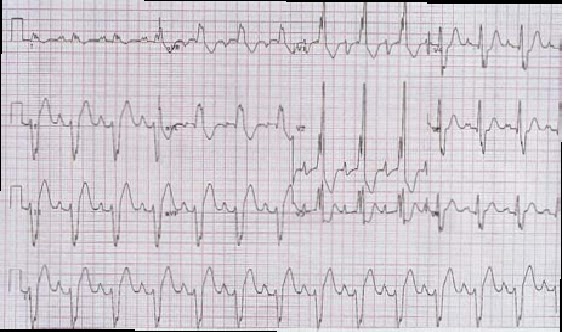
Study ECG ( For Postgraduate exams)


Regarding the ECG given above select five statements which are true from the list below:
a. Ventricular tachycardia.
b. Left axis deviation.
c. Voltage for left atrial enlargement.
d. LBBB.
e. Right axis deviation.
f. Voltage criteria for right ventricular hypertrophy.
g. RBBB.
h. Voltage criteria for right atrial enlargement.
i. Voltage criteria for left ventricular hypertrophy.
j. Dissociated P-waves.
k. Capture beats.
l. Delta waves.

Answer:

Sunday, October 9, 2016

Premature Ventricular Complexes (PVCs)

Which of the following statements about premature ventricular complexes (PVCs) is correct?

A) They are narrow electrocardiographic wave (QRS) complexes that are preceded by P waves.
B) In most cases, they disappear with exercise.
C) They are treated with type IC antiarrhythmics.
D) They may represent a risk for sudden death in healthy patients.
E) Caffeine use is not associated with PVCs

Answer: B ( In most cases, they disappear with exercise.)

Discussion: Premature ventricular complexes (PVCs) are abnormal ventricular beats that are characterized by wide QRS complexes, which are usually not preceded by P waves.


In patients with normal hearts, PVCs usually disappear with exercise. If the patient remains asymptomatic and there is no organic heart disease, no further treatment is necessary.

Friday, September 18, 2015

A 70 year Old Man With Hyperkalemia Secondary To Medication Side Effect

A 70 year old man with congestive cardiac failure and taking angiotensin converting enzyme inhibitor, spironolactone , digoxin and furosemide comes to the emergency department complaining of nausea and palpitations. He also mentions having not passed urine for last 24 hours. On admission an ECG was done which is shown below:

Note the tall peaked T waves.

What investigation should be performed next?

Answer: Serum potassium

Tall tented T-waves is an ECG feature of hyperkalaemia. In this case, the cause of hyperkalaemia is a combination of potassium-sparing drugs and obstructive uropathy.

Case Discussion:

Tuesday, July 21, 2015

A 40 Year Old Woman presents With Unsteady Gait And Recurrent Falls.

A 40-year-old woman presented with a five-day history of recurrent falls and an unsteady gait. She had a past history of a stroke causing a right-sided hemiparesis, which resolved spontaneously after a few days. The patient underwent intensive investigation following presentation with the stroke including carotid Doppler studies, transoesophageal echocardiography and CT scan
of the brain, which were normal.
The Ct Scan is shown below which is normal:

On examination she had a broad-based gait. There was evidence of dysdiadochokinesia in both upper limbs and abnormal heel–shin testing. The lower limb reflexes were brisk and the plantar response was extensor. The heart rate was 80 beats/min and regular. The blood pressure was 130/90 mmHg. Both heart sounds were normal.

Question :
What is the most probable diagnosis? ( Chose the one best from the options below)
a. Systemic lupus erythematosus with cerebralinvolvement.
b. Multiple sclerosis.
c. Multiple paradoxical emboli.
d. Friedreich’s ataxia.
e. Multiple cerebral metastases.


Answer:
b. Multiple sclerosis.

Explanation:

Monday, July 13, 2015

A 79 Year Old Female Presents With Worsening Confusion Over The Last 2 Days.

A 79-year-old female nursing home resident with moderate dementia presents for worsening confusion over the last 2 days. She just finished antibiotics for a urinary tract infection. Her temperature is 38.1◦C, pulse 110, respiratory rate 18, and blood pressure 118/60 mm Hg. She is disoriented and lethargic.
Examination of the heart, lungs, and abdomen is unremarkable.
Laboratory studies are as follows:
Na 165 mEq/L,
K 4.6 mEq/L,
Cl 118 mEq/L,
HCO3 28 mEq/L,
BUN 31 mg/dL,
Cr 1.1 mg/dL.
Urine specific gravity is >1.030 and urine osmolality is 700 mmol/kg(elevated reflecting reabsorption of free water)
 Her CBC is normal.

1. What will be the initial treatment for this patient?

A 15-Year Old Female With Right Upper Quadrant Abdominal Pain.

A 15-year-old Nigerian female was admitted with right upper quadrant abdominal pain. She had a history of sickle cell anemia, and had several admissions with hand and foot crises. During the last two admissions she complained of abdominal pain. She was born in England. She was the only child of a middle-class family.
On examination, she was distressed with pain. The temperature was 37.6°C (99.7°F). She was mildly jaundiced. The pulse was 105 beats/min, and blood pressure 100/75 mmHg. On examination of the respiratory system there was dullness to percussion on the anterior aspect of the right lung. Abdominal examination revealed tenderness in the right upper quadrant. The liver was palpable 3 cm below the costal margin.
Investigations are shown.


  • Hb 6 g/dl
  • WCC 12 109/l
  • Platelets 200 109/l
  • U&E Normal
  • AST 50 iu/l
  • Alkaline phosphatase 150 iu/l
  • Albumin 34 g/l
  • Urine osmolality 120 mOsm/kg
  • Urinalysis Urobilinogen +++, Blood 0, Protein 0 ,Bilirubin 0
  • Chest X-ray Opacification in the right upper zone


Practice Questions with Answers:

1. What is the cause of the abdominal pain?

Thursday, May 21, 2015

A 25 Year Old Man Presents With 3 Day History Of Chest Pain And Increasing Breathlessness.

A 25-year-old male presented to the Accident and Emergency Department with a three-day history of
pleuritic, lright-sided chest pain and increasing breathlessness. The symptoms started suddenly while he was playing football. He denied any chest trauma. Until then he had been fit and well, and there was no past medical history of note. His father died suddenly at the age of 32 years.
On examination he was thin and tall and distressed with pain. There was no pallor or cyanosis. He had a wide arm span. The heart rate was 100 beats/min and blood pressure 150/80 mmHg. The respiratory rate was 32/min. The trachea was deviated to the left. Percussion appeared to be reduced on the left side and was loud on the right side. The right lung base was dull to percussion. On auscultation of the lung fields breath sounds were absent on the right side and normal on the left side. Auscultation of the precordium demonstrated
quiet heart sounds and a soft mid-systolic murmur. The femoral pulses were easily palpable. The left calf was bruised and slightly tender. He attributed this to an injury sustained during the football match. The ECG revealed normal sinus rhythm.

1. Which test would you perform to reach a diagnosis?
Answer: Chest X ray.

The chest X ray is shown below:
The X ray Shows a large pneumothorax on the right side with a collapsed lung (arrows).

Thursday, May 14, 2015

A 45 year Old Man Taking Immunosuppressant Drugs presents With Jaundice And Fever

A 45 year old man with a 7 year history of rheumatoid arthritis presents with cough, high fever and a painless jaundice. Over the past 16 months his symptoms of joint pain were not controlled by NSAIDs alone and so he was switched to immunosuppressant drugs which helped in controlling his symptoms. Patient is a non smoker and consumes less than 2 units of alcohol per week.
On examination;

  •  patient looks unwell and deeply jaundiced.


  • He had a temperature of 102 F. 
  • Examination of the oral cavity shows yeast infection.


  • On auscultation of the lung fields there were coarse crackles at the right lung base. 
  • On abdominal examination spleen was palpable 3 cm below the left costal margin.
Investigations done are shown below: