Showing posts with label Pathology. Show all posts

Wednesday, November 1, 2017

Screening and Diagnosis of Celiac Diseases

Screening for celiac disease has been recommended for specific risk factors.

The anti-endomysium IgA antibody and anti-tissue transglutaminase IgA antibody tests are highly sensitive and specific in identifying individuals with celiac disease.

The anti-endomysium IgA antibody test is an immunofluorescent technique and is relatively expensive; interpretation is operator dependent and prone to errors so that it has largely been replaced by anti-tissue transglutaminase IgA antibody tests, which are simpler to perform and have similar sensitivity and specificity.

Anti-gliadin IgA and IgG and anti-reticulin IgA antibody tests are no longer recommended tests due to lack of specificity.

The anti-endomysium IgA and anti-tissue transglutaminase IgA antibody test can be falsely negative with IgA deficiency, which is associated with an increased incidence of celiac disease. Measurement of serum IgA concentration is mandatory to assure that false-negative results in IgA-deficient individuals are excluded. If celiac disease is suspected in patients with IgA deficiency, intestinal biopsy may be required. Because screening with antibodies may identify patients without documented celiac disease on biopsy, it is important to set the lower limit of antibody titers high enough to avoid false-positive results.

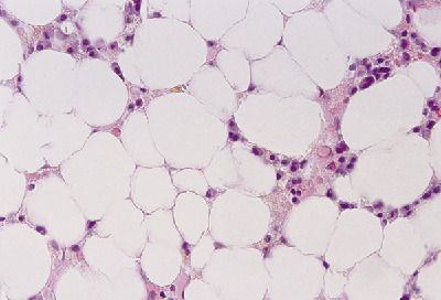
A. Normal
B. Celiac disease


Small Intestinal Biopsy.

Definitive diagnosis of celiac disease requires small intestinal biopsy, as none of the available serologic tests are 100% reliable. The characteristic histologic changes include partial or total villous atrophy, crypt elongation and decreased villous/crypt ratio, increased number of intraepithelial lymphocytes, intraepithelial lymphocyte mitotic index >0.2%, decreased height of epithelial cells, and loss of nuclear polarity. The mucosal involvement can be patchy, so multiple biopsies must be obtained.

Thursday, June 15, 2017

A sexually active 24-year-old woman presents to your office complaining of vaginal discharge.

A sexually active 24-year-old woman presents to your office complaining of vaginal discharge. Findings from a wet prep are pictured here.


The most likely diagnosis is
A) Yeast vaginitis
B) Gardnerella infection
C) Trichomonas infection
D) Gonorrhea
E) Chlamydia

Answer is

Monday, May 22, 2017

Introduction to Kaposi’s sarcoma

Kaposi’s sarcoma, a cancer of the lymphatic cell wall, affects tissues under the skin or mucous membranes that line the mouth, nose, and anus. In recent years, the incidence of Kaposi’s sarcoma has risen dramatically along with the incidence of human immunodeficiency virus (HIV) infection. It’s now the most common HIV-related cancer.
Kaposi’s sarcoma causes structural and functional damage. It progresses aggressively, involving the lymph nodes, the viscera and, possibly, GI structures.

Etiology
The exact cause of Kaposi’s sarcoma is unknown, but the disease may be related to immunosuppression. Genetic or hereditary predisposition is also suspected.

Signs and symptoms
The initial sign of Kaposi’s sarcoma is one or more obvious lesions in various shapes, sizes, and colors (ranging from red-brown to dark purple) that appear most commonly on the skin, buccal mucosa, hard and soft palates, lips, gums, tongue, tonsils, conjunctivae, and sclerae.
With advanced disease, the lesions may join, becoming one large plaque. Untreated lesions may appear as large, ulcerative masses.

                           Kaposi's Sarcoma Seen as a tumor on the roof of the mouth

                                                    Kaposi Sarcoma Affecting the skin
Other signs and symptoms include:
  • a history of HIV infection
  • pain (if the sarcoma advances beyond the early stages or if a lesion breaks down or impinges on nerves or organs)
  • edema from lymphatic obstruction
  • dyspnea (in cases of pulmonary involvement), wheezing, hypoventilation, and respiratory distress from bronchial blockage.
  • The most common extracutaneous sites are the lungs and GI tract (esophagus, oropharynx, and epiglottis).

Wednesday, May 10, 2017

APlastic and Hypoplastic Anemias


Aplastic and hypoplastic anemias result from injury to or destruction of stem cells in bone marrow or the bone marrow matrix, causing pancytopenia (anemia, granulocytopenia, thrombocytopenia) and bone marrow hypoplasia. Although often used interchangeably with other terms for bone marrow failure, aplastic anemias properly refer to pancytopenia resulting from the decreased functional capacity of a hypoplastic, fatty bone marrow.
These disorders generally produce fatal bleeding or infection, particularly when they’re idiopathic or stem from the use of chloramphenicol or from infectious hepatitis. Mortality for patients who have aplastic anemia with severe pancytopenia is 80% to 90%.

Causes
Aplastic anemias usually develop when damaged or destroyed stem cells inhibit red blood cell (RBC) production. Less commonly, they develop when damaged bone marrow microvasculature creates an unfavorable environment for cell growth and maturation. About half of such anemias result from drugs (antibiotics, anticonvulsants), toxic agents (such as benzene and chloramphenicol), or radiation. The rest may result from immunologic factors (unconfirmed), severe disease (especially hepatitis), or preleukemic and neoplastic infiltration of bone marrow.
Idiopathic anemias may be congenital and account for about 50% of all confirmed occurrences. Two such forms of aplastic anemia have been identified: congenital hypoplastic anemia (Blackfan-Diamond anemia), which develops between ages 2 months and 3 months, and Fanconi’s syndrome, which develops between birth and age 10.
With Fanconi’s syndrome, chromosomal abnormalities are typically associated with multiple congenital anomalies—such as dwarfism and hypoplasia of the kidneys and spleen. In the absence of a consistent familial or genetic history of aplastic anemia, researchers suspect that these congenital abnormalities result from an induced change in the development of the fetus.

Signs and symptoms
Signs and symptoms of aplastic anemias vary with the severity of pancytopenia but usually develop insidiously. These include progressive weakness and fatigue, shortness of breath, headache, pallor and, ultimately, tachycardia and heart failure. Thrombocytopenia leads to ecchymosis, petechiae, and hemorrhage, especially from the mucous membranes (nose, gums, rectum, and vagina) or into the retina or central nervous system. Neutropenia may lead to infection (with fever, oral and rectal ulcers, and sore throat) but without characteristic inflammation.

Wednesday, May 3, 2017

Corrosive Esophagitis

A 50 year old woman was seen in emergency after ingesting a chemical substance . Esophagogastrodudenoscopy was performed and the picture is shown below.


Initial esophagoscopy. (A) Middle esophagus shows whitish discoloration. (B) Distal esophagus shows exudates with easy touch bleeding.

The case was diagnosed as Corrosive Esophagitis 

Corrosive Esophagitis Case Discussion

Introduction
Inflammation and damage to the esophagus after ingestion of a caustic chemical is called corrosive or caustic esophagitis. Similar to a burn, this injury may be temporary or lead to permanent stricture (narrowing or stenosis) of the esophagus that requires corrective surgery.
Severe injury can quickly lead to esophageal perforation, mediastinitis, and death from infection, shock, and massive hemorrhage (due to aortic perforation).
Causes
The most common chemical injury to the esophagus follows the ingestion of lye or other strong alkalies; less commonly, injury follows the ingestion of strong acids. The type and amount of chemical ingested determine the severity and location of the damage.
In children, household chemical ingestion is accidental; in adults, it’s usually a suicide attempt or gesture. The chemical may damage only the mucosa or submucosa, or it may damage all layers of the esophagus.
Pathology
Esophageal tissue damage occurs in three phases: 
  1. in the acute phase, edema and inflammation; 
  2. in the latent phase, ulceration, exudation, and tissue sloughing; and
  3.  in the chronic phase, diffuse scarring.

Thursday, April 27, 2017

Salivary Gland Tumors

Different types of tumor may occur in the salivary gland and may be benign or malignant. A brief discription of the common tumors occuring in the salivary gland is given here:

1. Pleomorphic Adenoma
The pleomorphic adenoma- mixed salivary gland tumor- is a benign tumor, and the commonest salivary gland tumor. It is most commonly seen in the parotid gland and typically forms a mass in the lateral lobe, which slowly enlarges over many years, women in their forties are most often being affected.

Clinical Features
On palpation the tumor is firm, non tender and smooth or lobulated in texture. initially spherical and encapsulated, it may eventually spread more deeply, with the result that recurrence after resection is common. On oral examination the deep part of the gland may have pushed the tonsil and pillar of the fauces towards the midline.

Histology:
Histologically, it is highly variable in appearance, even within individual tumors. Classically it is biphasic and is characterized by an admixture of polygonal epithelial and spindle-shaped myoepithelial elements in a variable background stroma that may be mucoid, myxoid, cartilaginous or hyaline. Epithelial elements may be arranged in duct-like structures, sheets, clumps and/or interlacing strands and consist of polygonal, spindle or stellate-shaped cells (hence pleiomorphism). Areas of squamous metaplasia and epithelial pearls may be present. The tumor is not enveloped, but it is surrounded by a fibrous pseudocapsule of varying thickness. The tumor extends through normal glandular parenchyma in the form of finger-like pseudopodia, but this is not a sign of malignant transformation.
Pleomorphic adenoma consists of mixed epithelial (left) and mesenchymal cell components (right). The latter often exhibits myxofibrous appearance and in some instances shows chondromatous differentiation.
2. Adenolymphoma (Warthin’s Tumor)
It is a bening tumor and the second most common salivary tumor and is found exclusively in the parotid gland. It usually affects males in their fifties, forming a slow growing, painless swelling over the angle of the jaw, there is a high incidence of bilateral disease.

Monday, April 24, 2017

Insulinoma- A Pancreatic Islet Cell tumor


Introduction:
Insulinoma is a benign pancreatic islet cell tumor that produces excessive insulin and is derived from beta cells of the pancreas. The constant secretion of insulin from the tumor leads to hypoglycemia ( low blood sugar levels).

Incidence:
Insulinomas are rare tumors and mostly occur as a single small non cancerous growth is adults. People with genetic disorder known as multiple endocrine neoplasia type I are more prone to have this type of tumor. Its incidence is only 3-4 per million people making it one of the very rare tumors.

Clinical features:
  • Patients usually present with:
  • Recurrent headache
  • Lethargy
  • Anxiety
  • Behavioral changes
  • Blurred vision
  • Seizures
  • Coma if severe hypoglycemia occurs.
  • Confusion
  • Hunger
  • Sweating
  • Tremors

Friday, November 11, 2016

Pleural Effusion- A Brief Discussion

                                            CXR showing a large right pleural effusion

Pleural Effusion: Pleural effusion, sometimes referred to as “water on the lungs, is excess fluid that accumulates in the pleural cavity around the lungs.

The pleura are thin membranes that line the lungs and the inside of the chest cavity and act to lubricate and facilitate breathing. Normally, a small amount of fluid is present in the pleura.

Causes: Numerous medical conditions can cause pleural effusions. Some of the more common causes are: 
1.Transudative (watery fluid) pleural effusions:
  • Heart failure
  • Pulmonary embolism
  • Cirrhosis
  • Post open heart surgery
2. Exudative (protein-rich fluid) pleural effusions:
  • Pneumonia
  • Cancer
  • Pulmonary embolism
  • Kidney disease
  • Inflammatory disease

Thursday, November 10, 2016

Description Of Mouth Ulcers With Pictures For Better Learning

Introduction: Ulceration in the  mouth is a common occurrence and it is important for the physician to distinguish between the simple benign ulcers from those which are malignant.

1. Traumatic: The diagnosis is usually obvious with the clinical history. Patients may have ill fitting dentures or sharp teeth. The ulcer usually heals when the precipitating cause is removed.
 
                                               Traumatic ulcer

2. Aphthous Ulcers: Patients present with small, painful ulcers, usually inside the cheeks or lips. They heal in about one week but others may occur while the initial ulcer is healing. It may be secondary to stress, inflammatory bowel disease or an underlying debilitating disease,
On examination they are white, small, circular, deep , painful ulcers with surrounding erythema.
                                                      Aphthous Ulcer

3. Infective: May be due to bacteria ( Acute ulcerative gingivitis), Viral ( herpes simplex), or fungal (Candidiasis) .

In Acute ulcerative gingivitis (also known as Vincent's angina) patients present with swollen gums with small ulcers, which may spread to the buccal mucosa. There may be bleeding from the gums along with constitutional symptoms like fever and malaise. There is associated tender lymphadenopathy.
                                                    Acute ulcerative gingivitis

Candidiasis is common and is seen in patients with diabetes, immunosuppresed states or debilitating

Tuesday, October 11, 2016

Brief Discussion On Myeloproliferative Syndromes.

Myeloproliferative Syndromes is the name for a group of conditions that causes blood cells: RBCs, WBCs and platelets to grow abnormally in the bone marrow.

The three major myeloproliferative syndromes are:

1. Polycythemia Vera: It is the most common myleoproliferative disorder and is characterized by an increase in red blood cells (RBC) mass. massive splenomegaly and clinical manifestations related to increased blood viscosity. It also includes neurological manifestations like Vertigo, tinnitus, headache and visual disturbances. Increased RBC mass can also lead to thromboses that can causes complications like myocardial infarction, stroke and peripheral vascular disease.
Polycythemia vera must be distinguished from other causes of increased RBC mass, and this can be done by assaying serum erythropoietin levels. In polycythemia vera serum erythropoietin is low while in other causes  erythropoietin levels are high.
Bone marrow biopsy showing hypercellularity with trilineage growth (panmyelosis) with prominent erythroid, granulocytic, and megakaryocytic proliferation

Management: Patients are effectively managed with phlebotomy. Some patients may require splenectomy, and those with pruritits may benefit from psoralens and UV light.

2. Idiopathic Myelofibrosis: This rare entity is characterized by marrow fibrosis, myeloid metaplasia with extramedullary hematopoiesis and splenomegaly. Evaluation of blood smear reveals tear drop shaped RBC, nucleated RBC and some early granulocytic forms, including promyelocytes.

Thursday, September 29, 2016

A Case Of Urticaria (Hives)


A 45 Year old woman presents to her doctor complaining of itching and red patches on her skin of the legs, arms and abdomen. It first appeared 2 weeks back and has been increasing since then. She recalls having similar symptoms in the past at least 4 times . She relates it to start whenever she drinks citrus fruit juice. She was prescribed antihistamines and her symptoms gradually improved. She was advised to avoid food that trigger the allergy.

Case Discussion:

Hives also known as Urticaria is a skin reaction that presents as a red, raised, itchy skin rash. It appears and fades repeatedly. It usually happens due to an allergy.
Chronic hives is defined when the symptoms last more than six weeks or recur over months or years.

Pathophysiology; Whenever there is an allergic reaction triggered by a substance-allergen , body releases histamine . Histamine causes tiny blood vessels t leak fluid which accumulates in the skin causing the rash and swelling.

Causes : Females and young adults are more prone to utricaria. Each individual may develop symptoms due to a particular allergen to which his/her body responds and produces histamine.
Some of the common causes include:

Saturday, September 24, 2016

Effect Of Hyperuricaemia On Kidney.

The above picture shows uric acid kidney stone kept in the pathology lab. Let us briefly study the effects of Hyperuricaemia on kidney.

Causes Of Hyperuricaemia: High levels of urate in blood is known as hyperuricaemia. It may result from increased turnover (15%) or reduced excretion of urate (85%)
The causes are given below:

  • Drugs: cytotoxics, thiazides, loop diuretics,pyrazinamide
  • Increased cell turn over: Lymphoma, leukemia, psoariasisf, hemolysis, muscle death-rhabdomyolysis. 
  • Reduced excretion: Promary gout, chronic kidney disease, lead nephropathy, hyper parathyroidism, pre eclampsia
  • Others; Hypertension, Hyperlipedaemia 
  • Lesch-Nyhan syndrome - a disorder of purine synthesis
Hyperuricaemia and Renal Failure: Severe renal failure from any cause may be associated with hyperuricaemia, and rarely this may cause gout. Sometimes the relationship of cause and effect is reversed so that it is hyperuricaemia that causes the renal failure. This can occur in cytotoxic treatment (tumor lysis syndrome) and in muscle necrosis.

How Urate Causes Renal Failure?
Urate is poorly soluble in water so over excretion can lead to crystal precipitation. Renal failure occurs most commonly becauseurate precipitates in the renal tubules. This may occur at a plasma level > 1.19mmol/L. In some instances uretric obstruction from urate crystals may occur. This responds to retrograde uretric catheterization and lavage.

Monday, September 19, 2016

Acute Myeloid Leukemia

Acute Myeloid Leukemia (AML)

Also known as Acute myelogenous leukemia or Acute non lymphocytic leukemia

( The picture shows a blood smear in a patient with AML . Arrows indicate Aeur rods which are diagnostic of AML)

Introduction: AML is the rapidly progressive cancer of a group of white blood cells called myeloid cells ( which normally develop into various type of mature blood cells like red blood cells, white blood cells and platelets)

It is the commonest acute leukemia of adults, and its incidence increases with age. AML progresses rapidly and can cause death in few months if not treated.

Pathophysiology: AML is caused by damage to the DNA of developing cells in the bone marrow. The bone marrow produces immature cells that develop into leukemic white blood cells called myeloblasts. These abnormal cells build up and over crowd the bone marrow disturbing the function of healthy cells. In most cases the cause of theses DNA mutations is unknown.

Etiology: In most cases it is not known what leads to leukemia but radiation, exposure to certain chemicals and drugs, genetic disorders like Down syndrome and a previous cancer treatment are considered as risk factors for AML

Acute Lymphoblastic Leukemia

Acute Lymphoblastic Leukemia: ( ALL)


Also known as Acute Lymphocytic leukemia or Acute Lymphoid leukemia

Introduction: ALL is an acute cancer of the white blood cells affecting either B or T lymphocyte cell lines. The term acute comes from the fact that the disease progresses rapidly and creates immature blast cells with bone marrow failure and tissue infiltration.

Acute Lymphoblastic leukemia ALL is the most common type of cancer in children and luckily it has a good prognosis if treated . It is rare in adults.

Pathophysiology: In patients with ALL, there is overproduction of lymphobalsts in the bone marrow. These immature blast cells continuously multiply, and cause damage and death by inhibiting the production of normal red blood cells , white blood cells and platelets in the bone marrow. These malignant cells also infiltrate other organs causing damage.

Etiology: ALL is thought to develop from a combination of genetic susceptibility ( e.g with translocations amd gains and losses of whole chromosome) as well as an environmental trigger.
Ionizing radiations, eg X rays during pregnancy and Downs syndrome are important associations.

Clinical Features:
Patients usually present with recent onset of signs of marrow failure (pallor, fatigue, bleeding, fever, infection). Hepatosplenomegaly and adenopathy are common. males may have testicular enlargement reflecting leukemic involvement. Meningeal involvement may develop later.

Saturday, September 17, 2016

Studying The Erythrocyte (RBC) Morphology On Blood Smears

It is important for every medical student and doctor to learn the basic pathology like studying the morphology of normal and abnormal RBC's on blood smears. A brief description is given below.

Normal RBC Morphology: It is about 7.5 micrometer in diameter, roughly the size of a small lymphocyte. All the cells are of same size and color and have lightly colored area in the center.

Reticulocytes: are large, grey blue cells admixed with pink (on Wright's stain). Reticulocyte is actually an immature red blood cell without a nucleus. 


Anisocytosis: variation in RBC size, 
  • Normal MCV is 80-100 fl
  • large cells imply delay in erythroid precursor DNA synthesis caused by folate or B12 deficiency or as a side effect of medication. Macrocytes MCV > 100fl
  • small cells imply a defect in hemoglobin synthesis caused by iron deficiency or abnormal hemoglobin genes. Microcytes MCV < 80 fl 
RWD - the automated rec cell distribution width is a measure of anisocytosis.
Anisocytosis is a feature of most anemias


Poikilocytes: abnormal RBC shapes. They can be of many types and are described below:

Thursday, March 10, 2016

Pathogenesis In A Patient With Thrombotic Thrombocytopenic Purpura


A 38-year-old woman presents with a 3-day history of fever and confusion. She was previously healthy and is taking no medications. She has not had diarrhea or rectal bleeding. She has a temperature of 38°C (100.4°F) and a blood pressure of 145/85. Splenomegaly is absent. She has no petechiae but does have evidence of early digital gangrene of the right second finger. Except for confusion the neurological examination is normal. Her laboratory studies reveal the following:
Hemoglobin: 8.7
Platelet count: 25,000
Peripheral smear: numerous fragmented RBCs, few platelets
LDH 562 (normal <180)
Creatinine: 2.7
Liver enzymes: normal
Prothrombin time/PTT/fibrinogen level: normal

What is the most likely pathogenesis of her condition?

A. Disseminated intravascular coagulation
B. Antiplatelet antibodies
C. Failure to cleave von Willebrand factor multimers
D. Verotoxin-induced endothelial damage
E. Cirrhosis with sequestration of erythrocytes and platelets in the spleen

Answer And Discussion:

Tuesday, February 9, 2016

A Case Of Gastric Carcinoma

A 72-year-old Japanese immigrant was brought in by his family with complaints of difficulty in eating, vague abdominal pain, and weight loss. Endoscopy and biopsy confirmed gastric adenocarcinoma. Liver metastases were found on abdominal CT. The family and the patient chose only comfort measures and the patient died 6 months later.


Case Discussion:  

Introduction: Gastric cancer is a malignant neoplasm of the stomach, usually adenocarcinoma..

Epidemiology:  The median age at diagnosis is 70 years and median age at death from gastric cancer is 73 years.
• Stomach cancer occurs in 10.8 per 100,000 men and 5.4 per 100,000 women in a year. In 2008, the United States prevalence was 37,739 men and 28,271 women, with a lifetime risk of 0.88%.
• High rates of stomach cancer occur in Japan, China, Chile, and Ireland.

Pathophysiology: Eighty-five percent of stomach cancers are adenocarcinomas with 15% lymphomas and GI stromal tumors.
 Adenocarcinoma is further divided into two types:
~ Diffuse type—Characterized by absent cell cohesion, these tumors affect younger individuals infiltrating and thickening the stomach wall; the prognosis is poor. Several susceptibility genes
have been identified for this type of cancer.
~ Intestinal type—Characterized by adhesive cells forming tubular structures, these tumors frequently ulcerate.

Sunday, February 7, 2016

Folic Acid Deficiency Anemia


Essentials Of Diagnosis:

  • Macrocytic Anemia
  • Macro ovalocytes and hypersegmented neutrophils on peripheral blood smear
  • Normal serum vitamin B12 levels
  • Reduced folate levels in red blood cells or serum.
Introduction:
Folic acid is present in most fruits and vegetables ( especially citrus fruits and green leafy vegetables) and daily requirements of 50-100 mcg/d are usually met in the diet. 

Causes Of Folic Acid Deficiency: By far the most common cause of folic acid deficiency is inadequate dietary intake. Alcoholic or anorexic patients who do not eat fresh fruits and vegetables , and those who over cook their food are candidates for folate deficiency. Reduced folate absorption is rarely seen , since absorption occurs from the entire gastrointestinal tract. However certain drugs may lead to decreased folic acid absorption resulting in folate deficiency. 
Folic acid requirements are increased in pregnancy, hemolytic anemias and exfoliative skin diseases and the increased requirements may sometimes not be met by regular diet. The causes are summarized below: