Wednesday, November 2, 2016

A Case Of Normal-pressure hydrocephalus


A 75-year-old man is brought in to your office by his wife. She complains that he is not the same over the last 6 months. His memory is failing him, he has difficulty walking (especially when he initiates walking), and he is incontinent of urine. Which of the following is the most likely diagnosis based on his history?

Answer: Normal-pressure hydrocephalus (NPH)

Discussion:  Normal-pressure hydrocephalus(NPH) is a cause of dementia in the elderly. It may be
caused by previous insult to the brain, usually as a result of a subarachnoid hemorrhage or diffuse meningitis that presumably results in scarring of the arachnoid villi over the brain convexities where cerebrospinal fluid (CSF) absorption usually occurs. However, elderly NPH patients seldom have a
history of predisposing disease.

NPH classically consists of

  • dementia,
  • apraxia of gait, and 
  • incontinence 

Tuesday, November 1, 2016

Chest X ray Showing Left Thoracoplasty.

A 78-year-old man has been found collapsed on the street and is brought to the Emergency Department. He is cold and clammy. His ECG shows an acute anterior myocardial infarction. His CXR is shown below:


Question: What is the most likely cause for this patient’s radiological abnormality?

Answer: Left thoracoplasty

Discussion:

Atopic Dermatitis - Case Study.


This  7-month-old male child  is brought to clinic for a “rash all over.” Six weeks ago, his parents noticed him rubbing his legs against his crib and scratching his head frequently. They are concerned because they find blood on his sheets in the morning, and he has become increasingly irritable. He is eating and drinking normally.
His past medical history is unremarkable.
His father has sensitive skin and hay fever, but no one else in the family currently has a rash. He does not attend a daycare.
On skin exam, you find lichenified and erythematous patches of skin with fissures and bleeding on the ventral heels, dorsal feet, hands, and a few areas on the scalp. His cheeks are bright red with scale. His diaper area is uninvolved and there are no lesions in the web spaces of the hands and feet.

Q 1. Based on the description, which of the following is the most likely diagnosis?
A) Seborrheic dermatitis.
B) Atopic dermatitis (AD).
C) Scabies.
D) Tinea corpora.
E) Tinea versicolor.

Answer And Discussion

Plica Syndrome

A 15-year-old female athelete active in running competitions comes to your clinic complaining of knee pain over the medial knee joint. This pain is exacerbated by knee flexion and she notes popping and snapping when she stands from sitting. Your exam shows tenderness about 1 cm medial to the patella with palpable fullness in the area.
What is the most likely diagnosis is:
Answer: Plica Syndrome.

Case discussion:
Plica syndrome (also known as synovial plica syndrome) is a condition which occurs when a plica (an extension of the protective synovial capsule of the knee) becomes irritated, enlarged, or inflamed.


The presentation given in the case scenario is typical for plica syndrome. The plica is a synovial remnant that did not resorb properly during development. It can be irritated, usually chronically or sub acutely, especially in sports that require repeated flexion of the knee (rowing, cycling, running).
The inflammation is typically caused by the plica being caught on the femur, or pinched between the femur and the patella. The most common location of plica tissue is along the medial (inside) side of the knee.

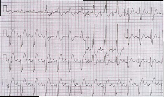
Study ECG ( For Postgraduate exams)


Regarding the ECG given above select five statements which are true from the list below:
a. Ventricular tachycardia.
b. Left axis deviation.
c. Voltage for left atrial enlargement.
d. LBBB.
e. Right axis deviation.
f. Voltage criteria for right ventricular hypertrophy.
g. RBBB.
h. Voltage criteria for right atrial enlargement.
i. Voltage criteria for left ventricular hypertrophy.
j. Dissociated P-waves.
k. Capture beats.
l. Delta waves.

Answer:

Diabetic Dermopathy

A 60-year-old woman with diabetes mellitus (DM) for the past 10 years began to notice reddish colored lesions on both anterior shins that turned brown over the past year. (Picture shown below: )


She reported having no pain with the hyperpigmented areas but does have foot pain secondary to neuropathy. The patient is diagnosed with diabetic dermopathy, and she begins working with her physician on achieving better control of her diabetes.

Case Discussion:

Diabetic dermopathy is a constellation of well-demarcated, hyperpigmented, atrophic depressions, macules, or papules located on the anterior surface of the lower legs that is usually found in patients with diabetes mellitus.(DM). It is the most common cutaneous marker of DM.

Diabetic dermopathy is found in 12.5% to 40% of patients with diabetes and most often in the elderly.

Etiology and Pathophysiology: The exact cause of diabetic dermopathy is unknown. Some of the suggested reasons include: