Wednesday, February 15, 2017

A 19 Year Old Woman With Fever And Sore Throat

A 19-yr-old woman presents with a severe sore throat, fever and malaise. She has marked cervical lymphadenopathy, gross splenomegaly and scattered petechiae on the soft palate, with enlarged tonsils covered by a confluent white exudate.


Her White cell count is mildly elevated, her serum ALT and AST concentrations are twice normal and her ALP is slightly elevated.
Which one of the following investigations is most likely to help guide your management?
A. FNA of a LN
B. HBsAg
C. CMV IgM
D. Heterophilic antibodies
E. HIV test

Answer:

Tuesday, February 14, 2017

Acute Right Ventricular Myocardial Infarction - ECG

Right Ventricular Myocardial Infarction. This ECG was obtained with right-sided lead placement.

ECG Findings
• ST elevation in right-sided V leads (V4R, V5R).
• ST elevation greater in lead III than lead II suggests RV MI.
• ST elevation in the normally obtained V1 also strongly suggests RV MI.

• Often associated with inferior MI and/or posterior MI.

ST elevation in V4R and V5R (arrows), with the V4 and V5 leads placed in their mirror-image locations on the right side of the chest. Any ST elevation seen in the right-sided precordial leads is significant.

Important Points: 
1. The smaller muscle mass of the right ventricle produces a less intense injury pattern that is

A 55-year-old woman complains of generalized fatigue, weakness and a rash.....

A 55-year-old woman complains of generalized fatigue, weakness, inability to climb stairs, arthralgias, and dysphagia.
Physical examination reveals definite proximal muscle weakness, a periorbital heliotrope rash, and skin findings associated with the hands (shown here).

The most likely diagnosis is
A) Lupus erythematosus
B) Sarcoidosis
C) Sjögren’s disease
D) Dermatomyositis
E) Polymyalgia rheumatica

Answer:

Chondrodermatitis nodularis helicis

Chondrodermatitis nodularis helicis (CNH) is a common and benign condition characterised by the development of a painful nodule on the ear.


Causes: It is thought to be caused by factors such as persistent pressure on the ear (e.g. secondary to sleep, headsets), trauma or cold. CNH is more common in men and with increasing age.

Clinical Presentation: The classic presentation of chondrodermatitis nodularis chronica helicis (CNH) is a middle-aged to elderly man with a spontaneously appearing painful nodule on the helix or antihelix. The nodule usually enlarges rapidly to its maximum size and remains stable. Onset may be precipitated by pressure, trauma, or cold. When asked, the patient usually admits to sleeping on the affected side.

On Examination: Nodules are firm, tender, well demarcated, and round to oval with a raised, rolled edge and central ulcer or crust. Removal of the crust often reveals a small channel. Color is similar to that of the surrounding skin, although a thin rim of erythema may be noted.

Pleural effusion On Chest X ray

A 28 years old male came to radiology department for X-ray chest with history of breathlessness on exertion since 10 days.
X-ray chest  shows a large pleural effusion on left side, the trachea and mediastinum are pushed to the right, right lung field is clear.

Pleural effusion is the accumulation of fluid in the pleural space, i.e. between the visceral and parietal layers of pleura.
The fluid may be transude, exudate, blood, chyle or rarely bile.
Pleural fluid casts a shadow of the density of water on the chest radiograph. The most dependent recess of the pleura is the posterior costophrenic angle. A small effusion will, therefore, tend to collect posteriorly; however, a lateral decubitus view is the most sensitive film to detect small quantity of free pleural effusion (as small as 50 ml). 100–200 ml of pleural fluid is required to be seen above the dome of the diaphragm on frontal chest radiograph. As more fluid is accumulated, a homogeneous opacity spreads upwards, obscuring the lung base. Typically this opacity has a fairly well-defined, concave upper edge , which is higher laterally and obscures the diaphragmatic shadow. Frequently the fluid will track into the pleural fissures.
A massive effusion may cause complete radiopacity of a hemithorax. The underlying lung will retract
towards its hilum, and the space occupying effect of the effusion will push the mediastinum towards the opposite side.

External Ear Injuries - Brief Description With Pictures.

Injuries to the external ear may be open or closed.

  • Blunt external ear trauma may cause a hematoma (otohematoma) of the pinna, which, if untreated, may result in cartilage necrosis and chronic scarring or further cartilage formation and permanent deformity (“cauliflower ear”). 
  • Open injuries include lacerations (with and without cartilage exposure) and avulsions

Pinna Hematoma. A hematoma has developed, characterized by swelling, discoloration, ecchymosis, and flocculence.  Immediate incision and drainage or aspiration is indicated, followed by an ear compression dressing.

Management: Pinna hematomas must undergo incision and drainage or large needle aspiration using sterile technique, followed by a pressure dressing to prevent reaccumulation of the hematoma.
This procedure may need to be repeated several times; hence, after Emergency department drainage, the patient is treated with antistaphylococcal antibiotics and referred to ENT or plastic surgery for follow- up in 24 hours. Lacerations must be carefully examined for cartilage involvement; if this is present, copious irrigation, closure, and postrepair oral antibiotics covering skin flora are indicated.

Saturday, February 11, 2017

A 19-yr-old woman Presents with Fever, Rash and Cough.....

A 19-yr-old woman presents with fever, rash and cough, and is pyrexial, tachycardic and tachypnoeic. She has a florid erythematous rash on her face, trunk and arms, with scattered whitish papular lesions on the buccal mucosa.


What is the most likely Dx?
A. Meningococcaemia
B. Rubella
C. Parvovirus B19
D. Secondary syphilis
E. Measles

Answer: E. Measles

Discussion: Adult measles is unusual except in non-immunised persons. The description of this Pt’s rash is classical: the rash is maculopapular, starts on the face and migrates caudally; Koplik’s spots are present in the mouth.

In contrast, meningococcaemia is a petechial/purpuric macular rash with no buccal lesions, and there is no typical migratory pattern.

Friday, February 10, 2017

Choking In An Adult - Management Algorithm

The management of choking is rightly taught as part of first aid. Recognition of the problem is the key to success. Clues include a person experiencing a sudden airway problem whilst eating, possibly combined with them clutching their neck.Victims with severe airway obstruction may be unable to speak or breathe and become unconscious.


Age-Related Macular Degeneration

Age-related macular degeneration increases in incidence with each decade over 50 and is evidenced by accumulation of either hard drusen (small, discrete, round, punctate nodules) or soft drusen (larger, pale yellow or gray, without discrete margins that may be confluent). Most patients with drusen have good vision, although there may be decreased visual acuity and distortion of vision. There may be associated pigmentary changes and atrophy of the retina. Vision may slowly deteriorate if atrophy occurs.
Patients with early or late degenerative changes of the macula are at risk of developing choroidal neovascularization (CNV), which is associated with distortion of vision, blind spots, and decreased visual acuity. Macular appearance may show dirty gray lesions, hemorrhage, retinal elevation, and
exudation.

Age-Related Macular Degeneration, Drusen. : Drusen are clustered in the center of the macula.

Management: Patients with drusen need ophthalmologic evaluation every 6 to 12 months or sooner if visual distortion or decreasing visual acuity develops. If a patient complains of deterioration of visual acuity or image distortion, prompt ophthalmic evaluation is warranted.

Important Clinical Points: 
1. Age-related macular degeneration is the leading cause of blindness in the United States in patients

Flail Chest In A Child Following An Accidental fall From The Rooftop.

A 8 years old child was brought to radiology department for X-ray chest following an accidental fall from the roof top.
His X ray is shown below:


X- Ray Description: X-ray chest (AP view of chest) shows 3rd to 8th rib fractures (white arrows) on left side, each at two places with encysted hemothorax (black arrow) and lung contusion (extends between the asterisks) with mediastinal pushed to the right side.

Diagnosis: Flail chest.

Clinical Discussion:
 Flail chest is a critical condition following major blunt chest trauma in which two or more

Wednesday, February 8, 2017

Bacterial Conjunctivitis - A Brief Introduction

Bacterial conjunctivitis is characterized by the acute onset of conjunctival injection and a thick yellow, white, or green mucopurulent drainage. Lid edema, erythema, and chemosis may also be seen.

Bacterial Conjunctivitis. Mucopurulent discharge, conjunctival injection, and lid swelling in a 10 year-old with H influenzae conjunctivitis.

  • Staphylococcus aureus is the most common causative bacteria.
  • Streptococcus pneumoniae and Haemophilus influenzae occur more frequently in children.


Bacterial Conjunctivitis. Mucopurulent discharge and conjunctival injection in an adult with conjunctivitis.

Hyperacute bacterial conjunctivitis, the most severe form of acute purulent conjunctivitis, is associated with N gonorrhoeae.
Symptoms are hyperacute in onset, and findings include a purulent, thick, copious discharge, eyelid swelling and tenderness, marked conjunctival hyperemia, chemosis, and preauricular adenopathy. The condition is serious and threatens sight because Neisseria species are capable of invading an intact corneal epithelium. Corneal findings include epithelial defects, marginal infiltrates, and an ulcerative keratitis that can progress to perforation.

Acid Base Balance - Mnemonic Charts


A 27 year Old Woman presents With Sudden Excessive Hair Loss


A 27-year-old woman , a mother of 2 children and who is now 6 months postpartum complains of excessive hair loss.
The most likely diagnosis is
A) Alopecia areata
B) Telogen effluvium
C) Trichotillomania
D) Tinea capitis
E) Hypothyroidism

The answer is

Acute Inferior-Posterior Myocardial Infarction - ECG

Acute Inferior-Posterior Myocardial Infarction: 


ECG Findings: 
• ST segment elevation in inferior leads (II, III, aVF)
• ST segment depressions in the anterior leads (V1-V3) and possibly high lateral leads (I, aVL)

Important Points: 
1. The right coronary artery supplies blood to the right ventricle, the sinoatrial (SA) node, the inferior portions of the left ventricle, and usually to the posterior portion of the left ventricle and the atrioventricular (AV) node.
2. Infarctions involving the SA node may produce sinus dysrhythmias including tachycardias, bradycardias, and sinus arrest.

Different Types Of Tremors - Chart

Tuesday, February 7, 2017

Nevus Simplex Or A Salmon Patch - A Vascular Lesion Of Infancy

Nevus simplex (salmon patch) is the most common vascular lesion in infancy, present in about 40% to 60% of newborns. It appears as a blanching, slightly pink-red macule or patch most commonly on the nape of the neck, the glabella, mid-forehead, or upper eyelids. Lesions generally fade over the first 2 years of life but may become more prominent with crying or straining.

                                 Newborn with characteristic salmon patches over his face.

Important Points:
1. Salmon patches are composed of ectatic dermal capillaries.
2. Salmon patches appear symmetrically and cross the midline in contrast to the unilateral distribution of a port-wine stain.
3. When seen on the nape of the neck, this lesion is referred to as a stork bite  (see picture below) or as an angel’s kiss when appearing on the forehead.

Monday, February 6, 2017

Treatment Algorithm And A Brief Description Of Anaphylaxis


Anaphylaxis is a generalized immunological condition of sudden onset, which develops after exposure to a foreign substance.

Pathophysiology: The mechanism may:
• Involve an IgE-mediated reaction to a foreign protein (stings, foods, streptokinase), or to a protein–hapten conjugate (antibiotics) to which the patient has previously been exposed.
• Be complement mediated (human proteins eg G -globulin, blood products).
• Be unknown (aspirin, ‘idiopathic’).
Irrespective of the mechanism, mast cells and basophils release mediators (eg histamine, prostaglandins, thromboxanes, platelet activating factors, leukotrienes) producing clinical manifestations.
Angio-oedema caused by ACE inhibitors and hereditary angio-oedema may present in a similar way
to anaphylaxis. Hereditary angio-oedema is not usually accompanied by urticaria and is treated with C1 esterase inhibitor.

Common causes: include:

Acute Anterior Myocardial Infarction - ECG


ECG Findings
• ST segment elevation in the anterior precordial leads.
• V1-V4: Anteroseptal injury.
• V3-V4: Anterior injury.
• V3-V6: Anterolateral injury. Leads I and aVL may also be involved, especially if the circumflex artery is affected (high lateral injury).
• Reciprocal ST segment depressions are often present in the inferior leads (II, III, aVF).

Important Points To Remember: 
1. The left anterior descending artery supplies blood to the anterior and lateral left ventricle and ventricular septum.
2. Normal R-wave progression (increasing upward amplitude with R wave > S wave at V3 or V4) may be interrupted.

Sunday, February 5, 2017

Introduction To Chilblains (Pernio)


Chilblains (sometimes referred to as pernio) describes a number of symptoms which occur in the peripheries (e.g. toes, fingers, earlobes) in response to the cold. It is thought to be caused by an abnormal vascular response to cold exposure.

Clinical Signs And Symptoms: 
  • The areas most affected are the toes, fingers, earlobes, nose.
  • Blistering of affected area
  • Burning and itching sensation in extremities
  • Dermatitis in extremities
  • Digital ulceration (severe cases only)
  • Erythema (blanchable redness of the skin)
  • Pain in affected area
  • Skin discoloration, red to dark blue
  • Chilblains usually heal within 7–14 days.
Risk factors: Factors that may increase risk of  developing chilblains include:

Depressed Skull Fracture

Depressed skull fractures typically occur when a large force is applied over a small area. They are classified as open if the skin above them is lacerated. Abrasions, contusions, and hematomas may also be present over the fracture site.
The patient’s mental status is dependent upon the degree of underlying brain injury. Direct trauma can cause abrasions, contusions, hematomas, and lacerations without an underlying depressed skull fracture.  Evidence of other injuries such as a basilar fracture, facial fractures, or cervical spinal injuries may also be present.

                                            CT demonstrating depressed skull fracture. 

Management: Explore all scalp lacerations to exclude a depressed fracture. CT should be performed in all suspected depressed skull fractures to determine the extent of underlying brain injury.

  • Depressed skull fractures require immediate neurosurgical consultation. 
  • Treat open fractures with antibiotics and tetanus prophylaxis as indicated. 
  • The decision to observe or operate immediately is made by the neurosurgeon. 

COPD ( Emphysema And Chronic Bronchitis) Mnemonic


Metastasis - Detected On Radiology

A 58 years old male came to his physician complaining of chest pain  for about 6 months. The pain was initially mild but now has gradually increased in severity. The patient mentions feeling tired all the time with no energy and unintentional weight loss.
He was sent for a chest X ray which is shown below:

X-ray chest shows a large (8 × 6 cm) well-defined mass lesion abutting the left lower chest wall with broad base towards the chest wall with partial destruction of lateral aspect of 4th rib on the left.

It was suspected to be a malignant lesion. CT chest was advised which is shown below:

Contrast CT chest (Figs A to D) was done which shows moderately enhancing metastatic bone lesion which is rounded and well-defined having a large soft tissue component from the left 4th rib, and right 10th rib (Figs B and C) laterally, the ribs are partially destroyed, few small scattered calcific densities are seen in the lesions. There is a single iso to hypodense lesion seen in liver measuring 5 × 4 cm. The lesion shows moderate heterogeneous post-contrast enhancement (Fig. D).

Diagnosis: Metastasis to ribs and liver

Case Discussion: Primary tumors which originate in other organs and involve the skeletal structures of the body either by hematogenous, lymphatic route or by direct invasion are called metastasis.

Saturday, February 4, 2017

Introduction To Erythema multiforme

Definition: Erythema multiforme (EM) is an acute, self-limited, and sometimes recurring skin condition that is considered to be a type IV hypersensitivity reaction.
Etiology: It occurs in response to medicines, infections, or illness

  • Herpes simplex virus (HSV; frequently labialis) is strongly associated but may not be clinically apparent. Other viruses, bacteria (M pneumoniae, Chlamydia, Salmonella, Mycobacterium), and fungi (Histoplasma capsulatum, dermatophytes)
  • are also associated. 
  • Medications account for <10%; NSAIDs, sulfonamides, antiepileptics, allopurinol, and antibiotics are
  • responsible for the majority. 
  • Physical factors such as trauma, ultraviolet light exposure, and cold have been reported to elicit EM.

Clinical Features: 

Erythema Multiforme. Symmetric distribution of targetoid macules and plaques. The dusky central zone is more obvious on the left waistline lesions.

  • Erythema multiforme (EM) begins with symmetric, erythematous, sharply defined extremity or trunk macules, and evolves into a “targetoid” or “bull’s eye” morphology (a flat, dusky, central area with two concentric, erythematous rings).
  • Bullae may appear in the central dusky area (bullous EM).

Brachial Plexus - Anatomy


Shoulder Dislocation - A Brief DIscussion

Anterior shoulder dislocations are the most common and frequently caused by falling with the arm externally rotated and abducted.

Clinical Presentation: Patients present with the affected extremity held in adduction and internal rotation. Often, they complain of shoulder pain, refuse to move the affected arm, and may support the dislocated shoulder with the other arm. The acromion becomes prominent with loss of the rounded contour of the deltoid. A neurovascular examination of the upper extremity should be
performed to rule out associated injury, most commonly of the axillary nerve (sensation over the deltoid) and of the musculocutaneous nerve (sensation on the anterolateral forearm). Vascular injuries have rarely been reported to occur.

Diagnosis: Standard radiographic examination to evaluate for associated fracture should include AP and either axillary lateral or scapular views.

Anterior Shoulder Dislocation. Radiographic evaluation demonstrates that the humeral head is not in the glenoid fossa but is located anterior and inferior to it.


Posterior shoulder dislocations are commonly missed because of subtle radiographic findings.

Thursday, February 2, 2017

A 35 Year Old Patient With Progressive Hearing Loss

A 35-year-old presents with unilateral hearing loss that has been gradual but progressive over the last 6 months. Otoscopy reveals a Cholesteatoma.


Appropriate treatment of the above condition consists of
A) Prolonged antibiotics for up to 4 weeks
B) Decongestant and antihistamine administration
C) Corticosteroid treatment for 2 weeks
D) Hearing aid amplification
E) Tympanomastoidectomy

Answer:

Cherry Hemangiomas (Campbell De Morgan spots Or Senile Angiomas)


Cherry hemangiomas 
also known as Campbell De Morgan spots or senile angiomas are the most common cutaneous vascular proliferations. They are often widespread and appear as tiny cherry-red papules or macules. Longstanding lesions enlarge slowly over time and take on the appearance of a dome topped with cherry-red to deep-purple papules.

They are more common with advancing age and affect men and women equally.

Clinical Features
  • erythematous, papular lesions
  • typically 1-3 mm in size
  • nonblanching
  • not found on the mucous membranes
Causes: The exact cause of cherry angiomas is unknown, but there may be a genetic factor that

Cellulitis - A Brief Discussion

Cellulitis is a common infection of the skin or subcutaneous tissues with characteristic findings of:
  • erythema with poorly defined borders, 
  • edema, warmth, pain, and limitation of movement.
  • Fever and constitutional symptoms may be present and are commonly associated withm bacteremia. 
     Cellulitis of the right lower extremity characterized by sharply demarcated erythema an edema.


Predisposing factors include:


  • trauma, 
  • lymphatic or venous stasis, 
  • immunodeficiency (including diabetes mellitus), and 
  • foreign bodies.

Common etiologic organisms include:

  • group A β-hemolytic Streptococcus 
  • Staphylococcus aureus in nonintertriginous skin, and  gram-negative organisms or mixed flora in intertriginous skin and ulcerations. 
  • In immunocompromised hosts, Escherichia coli, Klebsiella species, Enterobacter species, and Pseudomonas aeruginosa are common. 
  • In recent years, there has been a dramatic increase in the incidence of community- acquire methicillin-resistant S aureus (CA-MRSA), particularly in cellulitis associate with a cutaneous abscess.

Wednesday, February 1, 2017

A 19 Year Old Male With Cough, headache and Malaise...

A 19-yr -old army cadet is admitted to hospital with cough, headaches and malaise. He has a temperature of 38°C. His blood count and renal and liver function are normal. Cold agglutinins
are positive. A CXR shows bibasal shadowing.


What is the most likely Dx?

A. Legionella pneumonia
B. Viral pneumonia
C. Q fever
D. Klebsiella pneumonia
E. Mycoplasma pneumonia

Answer:

A 3 Year Old Child With A Mass On The Chest

A 3 years old child was seen by the pediatrician for a large soft tissue mass on right side of chest.

Photograph (A) of the child shows the soft mass (arrow), on X-ray chest (B) the soft tissue lymph nodal mass lies outside the thoracic cage in right hemithorax.

Diagnosis: Lymph node mass.

Discussion: On X-ray chest the lung fields are clear, on X-ray chest it is important to look for any lesion in the soft tissue over the entire available film. It may be in the form of enlarged thyroid gland, cervical or axillary lymph nodes, neurofibroma, surgical emphysema, a lesion in the breast or one
of the breasts might have been surgically excised. Ultrasound can be useful in investigating nodal masses particularly as an aid to biopsy.

Acromioclavicular Joint Seperation

Injury to the acromioclavicular (AC) joint usually results from an impact on the superior aspect of the acromion.

The classification system for AC joint injuries includes six types.

  • A type I injury is equivalent to a stretching of the AC ligament. 
  • A type II injury consists of tearing of the AC ligaments and stretching of the coracoclavicular ligaments. 
  • Complete disruption of the AC and coracoclavicular ligaments is seen in types III to VI.


Clinical Features: Patients complain of pain at the AC joint and will actively splint the injured shoulder. Ecchymosis may be present; however, an obvious deformity is not always seen. There is significant tenderness upon palpation of the AC joint.

Diagnosis: Standard radiographs should include anteroposterior (AP) and axillary lateral views of the shoulder.

Testicular Torsion

 A young male , age 16 years presents to the emergency with the complain of the sudden onset of pain in one testicle, followed by swelling of the affected testicle, reddening of the overlying scrotal skin, lower abdominal pain, nausea, and vomiting.
An examination reveals a swollen, tender, retracted testicle that often lies in the horizontal plane (bell-clapper deformity).

A diagnosis of Testicular torsion was made and the patient was prepared for immediate surgery.

Case Discussion:


Testicular torsion is one of the urologic emergencies and is very painful on presentation.

Testicular torsion occurs when a testicle rotates, twisting the spermatic cord that brings blood to the scrotum. The reduced blood flow causes sudden and often severe pain and swelling.

Testicular torsion is most common between ages 12 and 16, but it can occur at any age, even before birth.

Traumatic Asphyxia - Brief Discussion

Traumatic Asphyxia:  Traumatic asphyxia, also known as  Perthe's syndrome, is a medical emergency caused by an intense compression of the thoracic cavity, causing venous back-flow from the right side of the heart into the veins of the neck and the brain

The clinical findings of traumatic asphyxia are due to a sudden increase in intrathoracic pressure against a closed glottis. The elevated pressure is transmitted to the veins, venules, and capillaries of the head, neck, extremities, and upper torso, resulting in capillary rupture.

Strangulation and hanging are common mechanisms. Survivors demonstrate plethora, ecchymoses, petechiae, and subconjunctival and retinal hemorrhages.

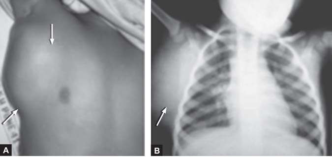
Body showing  dark purple Tardieu spots in dependent areas, due to ruptured capillaries in a person who suffered from Traumatic Asphayxia
Severe injuries may produce central nervous system injury with blindness, seizures, posturing, and paraplegia and even death.

Management: Treatment is supportive, with attention to other concurrent injuries. Long-term

Understanding Axis Deviation On ECG