Friday, November 3, 2017

Fungal Infection of Nails

Fungal infection of the nails has been classified into four types :

1. White superficial onychomycosis presents with diffuse or speckled white discoloration of the surface of the toenails. It is caused primarily by Trichophyton mentagrophytes, which invades the nail plate. The organism may be scraped off the nail plate with a blade, but treatment is best accomplished by the addition of a topical azole antifungal agent.


2. Distal subungual onychomycosis presents with foci of onycholysis under the distal nail plate or along the lateral nail groove, followed by development of hyperkeratosis and yellow-brown discoloration. The process extends proximally, resulting in nail plate thicken and separation from the nail bed. Trichophyton rubrum and, occasionally, T. mentagrophytes infect the toenails; fingernail disease is almost exclusively due to T. rubrum, which may be associated with superficial scaling of the plantar surface of the feet and often of one hand. These dermatophytes are found most readily at the most proximal area of the nail bed or adjacent ventral portion of the nail plates that are involved. Topical therapies such as ciclopirox 8% lacquer may be effective for solitary nail infection. Because of their long half-life in the nail, terbinafine or itraconazole may be effective when given as pulse therapy (1 wk of each mo for 3–4 mo). Either agent is superior to griseofulvin, fluconazole, orketoconazole. The risks, the most concerning of which is hepatic toxicity, and costs of oral therapy must be weighed carefully against the benefits of treatment for a condition that generally causes only cosmetic problems.

Wednesday, November 1, 2017

Screening and Diagnosis of Celiac Diseases

Screening for celiac disease has been recommended for specific risk factors.

The anti-endomysium IgA antibody and anti-tissue transglutaminase IgA antibody tests are highly sensitive and specific in identifying individuals with celiac disease.

The anti-endomysium IgA antibody test is an immunofluorescent technique and is relatively expensive; interpretation is operator dependent and prone to errors so that it has largely been replaced by anti-tissue transglutaminase IgA antibody tests, which are simpler to perform and have similar sensitivity and specificity.

Anti-gliadin IgA and IgG and anti-reticulin IgA antibody tests are no longer recommended tests due to lack of specificity.

The anti-endomysium IgA and anti-tissue transglutaminase IgA antibody test can be falsely negative with IgA deficiency, which is associated with an increased incidence of celiac disease. Measurement of serum IgA concentration is mandatory to assure that false-negative results in IgA-deficient individuals are excluded. If celiac disease is suspected in patients with IgA deficiency, intestinal biopsy may be required. Because screening with antibodies may identify patients without documented celiac disease on biopsy, it is important to set the lower limit of antibody titers high enough to avoid false-positive results.

A. Normal
B. Celiac disease


Small Intestinal Biopsy.

Definitive diagnosis of celiac disease requires small intestinal biopsy, as none of the available serologic tests are 100% reliable. The characteristic histologic changes include partial or total villous atrophy, crypt elongation and decreased villous/crypt ratio, increased number of intraepithelial lymphocytes, intraepithelial lymphocyte mitotic index >0.2%, decreased height of epithelial cells, and loss of nuclear polarity. The mucosal involvement can be patchy, so multiple biopsies must be obtained.

Monday, October 30, 2017

Some Limb Deformities in a Newborn

1. Metatarsus adductus. In metatarsus adductus, the forefoot is deviated medially and is slightly supinated. In the normal foot, a line drawn through the hindfoot will pass between the second and third toe. With metatarsus adductus, this line will pass lateral to the third toe. The prognosis is excellent, with most cases of metatarsus adductus resolving spontaneously.


2. Calcaneovalgus. Calcaneovalgus deformity of the foot is commonly associated with lateral tibial torsion. The forefoot is abducted, and the ankle is severely dorsiflexed to where the foot folds against the anterolateral surface of the tibia. The deformity is usually flexible, and the foot can be passively placed in the normal position. Calcaneovalgus deformity of the foot usually resolves spontaneously.


3. Talipes equinovarus. Talipes equinovarus, or club foot, has an incidence of approximately 1.5 in 1,000. Fifty percent of the time, the condition is bilateral. It can be associated with other conditions such as spina bifida and arthrogryposis. The foot turns inward and downward and remains tight in this position. Talipes equinovarus requires immediate evaluation since the timing of corrective casting, if necessary, can affect optimal outcome.

Tuesday, August 22, 2017

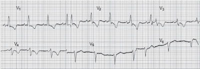
A 55 Year Old Man Presents To Emergency With Chest Pain For 6 Hours...

The ECG shown below was recorded in the A & E department from a 55-year-old man who had had chest pain at rest for 6 hours. There were no abnormal physical findings.
What does the trace show, and how would you manage him?


The ECG shows:
• Sinus rhythm
• Normal axis
• Normal QRS complexes
• ST segment depression - horizontal in leads V3-V4, downward-sloping in leads I, VL, V5-V6

Monday, July 31, 2017

Important Clinical Findings In The Tympanic Membrane and Middle Ear

“Chalk” patches. White areas of tympanosclerosis are common findings on examination of the ear drum. They are of little significance in themselves, and the hearing is often normal.

A past history of otorrhea in childhood or grommet insertion is usual. Chalk patches do occur with no
apparent past otitis media. Extensive tympanosclerosis with a rigid drum is a sequela of past otitis media, and the ossicles, too, may be fixed or noncontinuous.

Scarring of the drum. A gossamer-thin membrane can be seen to close this previously well-defined central perforation (arrow).


At first sight with the auriscope, a central perforation would appear to be the diagnosis; more careful
examination with a pneumatic otoscope will show that this thin membrane moves and seals the defect, giving reassurance that the drum is intact.

Scarring of the drum with retraction onto the round window, promontory, and incus is also evidence of past otitis media.

It is sometimes difficult to be sure whether this type of drum is intact; a thin layer of epithelium indrawn onto the middle-ear structures may seal the middle ear, and examination with the operating
microscope may be necessary to be certain of an intact drum.

Scarred tympanic membrane. A scarred tympanic membrane in which the drum has become atelectatic and indrawn onto the long process of the incus and promontory.


A retracted tympanic membrane which is thin and indrawn onto the long process of the incus (a), head of the stapes (b), promontary (c), and round window (d).

The stapedius tendon is also seen in this panoramic view obtained with a fiberoptic endoscope.

Creatinine Kinase (CK-MB) Learning Flash Card


Monday, July 17, 2017

Onychomycosis and Diabetes: Patient Case Challenges

Onychomycosis is a common infection that can lead to potentially severe complications in patients who have impaired circulation, peripheral neuropathy, or are immunocompromised. This infection is particularly common in patients with diabetes, many of whom have one or more of these conditions. Onychomycosis treatment reduces risks, but is complicated by the potential for drug-drug interactions between systemic antifungals and the large number of medications often taken by these patients. Several topical therapies are available, including two newer agents that became available in 2014, increasing the potential for individualized treatment selection.

The following case-based activity will test your ability to select appropriate therapies in patients with type 2 diabetes and onychomycosis, while bringing you up to date on risks and management of toenail infections in this vulnerable patient population.

Case :
Albert is a 55-year-old man with type 2 diabetes who presents to your office with thickened, elongated, yellowed toenails (picture shown below). He reports that he first noticed the changes in his toenails about 3 months ago, at which time most of his nails were already involved. Albert was diagnosed with type 2 diabetes 4 years ago, and his blood sugar is currently poorly controlled on metformin alone. He has elevated triglyceride levels for which he is taking a low dose of simvastatin. He is clinically obese, which may have contributed to the fact that he did not notice his toenail infection until it was well entrenched.


On clinical examination, you observe that all of Albert’s toenails are yellow in color and 6 have white lateral streaks. Two nails show involvement extending proximally to the lunula. Several nails, including both great toenails, show subungual hyperkeratosis that is greater than 2 mm thick and two are onycholytic. You estimate that at least 75% of the overall area of Albert’s toenails is infected, with involvement seen in all 10 toenails.

Which of the following clinical characteristics is a risk factor for onychomycosis in patients with diabetes?
A. Female sex
B. Neuropathy
C. Renal dysfunction
D. Younger age

Answer: B. Neuropathy
Discussion: Patients with diabetes can develop peripheral neuropathy, which contributes to the risk of developing onychomycosis, perhaps because this condition impedes proper foot care and allows unrecognized trauma to the nail. 
Men with diabetes are approximately 3 times more likely to develop onychomycosis compared with women with diabetes, and the prevalence of onychomycosis increases with age. Onychomycosis does not seem to be linked to the presence or absence of renal dysfunction in patients with diabetes.

Onychomycosis and Diabetes
Onychomycosis is highly prevalent in patients with diabetes. A North American study of 550 consecutive patients with diabetes found abnormal-appearing toenails in 46% of patients and mycological evidence of onychomycosis in 26% of patients. After adjusting for age, the researchers calculated a 34.9% prevalence in the overall US diabetes population, an estimate that is similar in other studies. Onychomycosis was 2.77 times more likely in patients with diabetes compared with individuals without diabetes. Prevalence in the one-third of patients with type 1 diabetes in this study was lower but still elevated compared with individuals without diabetes, with an odds ratio of 1.69. Onychomycosis was also 3 times more likely in men compared with women with diabetes.

Chilaiditi Sign On Chest X ray

The X ray shown below is of an asymptomatic person. 


Description Of Chilaiditi Sign
Chilaiditi described this normal variant in 1911 where the transverse colon is interposed between the right hemidiaphragm and the liver.
Its prevalence is thought to be 0.025%.

Occasional reports describe patients with Chilaiditi’s syndrome where patients complain of intermittent abdominal pain requiring laparotomy to rule out other causes of peritonism, e.g. perforated ulcer, ruptured appendix.

Atrial Fibrillation - ECG and Learning Questions/Answers


ECG Criteria
• P wave: Absent (P may be replaced by fibrillary f wave).
• Rhythm: Irregularly irregular (R-R interval is irregular). (Atrial rate is very high and ventricular rate is less).

According to the rate, atrial fibrillation may be of 2 types:
• Fast atrial fibrillation: Heart rate >100 beats/min.
• Slow atrial fibrillation: Heart rate <100 beats/min.

Q. What is atrial fibrillation?
Ans. It is an arrhythmia where atria beat rapidly, chaotically and ineffectively, while the ventricles respond at irregular intervals, producing the characteristic irregularly irregular pulse. Any conditions causing raised atrial pressure, increased atrial muscle mass, atrial fibrosis, inflammation and infiltration of the atrium can cause atrial fibrillation.

Q. What are the types of atrial fibrillation?
Ans. There are 3 types of atrial fibrillation:
• Paroxysmal: Discrete self-limiting episodes. May be persistent if underlying disease progresses.
• Persistent: Prolonged episode that can be terminated by electrical or chemical cardioversion.
• Permanent: Sinus rhythm cannot be restored.

Wednesday, July 12, 2017

Keloid Scars - A Brief Discussion


Keloid scars are tumor like lesions that arise from the connective tissue of a scar and extend beyond the dimensions of the original wound

Predisposing factors

  • ethnicity: more common in people with dark skin
  • occur more commonly in young adults, rare in the elderly
Pathophysiology: When skin is injured, fibrous tissue, called scar tissue, forms over the wound to repair and protect the injury. In some cases, scar tissue grows excessively, forming smooth, hard growths called keloids. Keloids can be much larger than the original wound.


Common Sites (in order of decreasing frequency):

  • sternum, 
  • shoulder,
  • neck,
  • face, 
  • extensor surface of limbs, 
  • trunk
Keloids should not be confused with hypertrophic scars, which are raised scars that do not grow beyond the boundaries of the original wound.

Tuesday, July 11, 2017

A 26-year-old woman, who has complained of palpitations in the past, presents to the emergency department ....

A 26-year-old woman, who has complained of palpitations in the past, is admitted via the A & E department with palpitations. Her ECG is shown below:


What does the ECG show and what should you do?

The ECG shows:
• Narrow-complex tachycardia, rate about 200/min
• No P waves visible
• Normal axis
• Regular QRS complexes
• Normal QRS complexes, ST segments and T waves

Clinical interpretation
This is a supraventricular tachycardia, and since no P waves are visible this is a junctional, or
atrioventricular nodal, tachycardia.

Diagnosis: Junctional (atrioventricular nodal re-entry) tachycardia.

What to do
Junctional tachycardia is the commonest form of paroxysmal tachycardia in young people, and

A 26 year old man with lesions on his body...

A 26-year-old man is seen for an upper respiratory infection. After removing his shirt, you notice the lesions shown in the picture below. He reports that his father has similar lesions.


The differential diagnosis should include
A) Hypothyroidism
B) Addison’s disease
C) Multiple sclerosis
D) Neurofibromatosis
E) Gardner’s syndrome

Answer:

Ashman Phenomenon - ECG


ECG Findings
• Aberrant ventricular conduction, usually with RBBB pattern.
• Altered durations of the refractory period of the bundle branch or ventricular tissue are present, commonly due to atrial fibrillation, atrial ectopy, and atrial tachycardia

Points to Remember:

1. After depolarization, tissue repolarizes during its refractory period. Refractory period changes with the preceding cardiac cycle, with longer R-R intervals producing longer refractory periods and shorter R-R intervals producing shorter refractory periods.

2. A longer R-R interval lengthens the following refractory period. When an early or premature (ectopic) depolarization reaches the ventricular conduction system before it has completely repolarized, aberrant conduction may occur and be manifest on the ECG with a bundle branch block (BBB) pattern.

3. Ashman phenomenon most commonly appears with an RBBB pattern, since the right bundle has a longer refractory period than the left bundle.

4. Ashman phenomenon is often seen in atrial fibrillation, when a long R-R interval is followed by a much shorter R-R interval.

Pulmonary Alveolar Microlithiasis - Chest X Ray

A 39 years old female came to radiology department for X-ray chest with history of dyspnea on exertion. The X ray is shown as below:


Radiological Findings on the X ray Chest: X-ray chest (in the picture above) shows that the lung fields are diffusely occupied by discrete high density opacities resembling grains of sands. In spite of
superimposition or summations of shadows the individual deposits are identifiable and measure about a 1 mm in diameter thick and streaky. The opacity appears confluent showing the lungs as almost white with obliteration of the mediastinal and diaphragmatic contours and pulmonary vascular marking are indistinct. The density is greater over the lower than the upper zone.

Comments and Explanation: The characteristic finding on the chest radiograph is that of a fine, dense stippling. This is a well-defined, nodular infiltrate which involves both lungs, and may be so extensive as to cause opaque lungs with relative lucency of the mediastinal structures, diaphragm and pleura. There is widespread parenchymal calcification.
In addition to the finding seen, other findings that may be seen include bullae in the lung apices, a zone of increased lucency between lung parenchyma and the ribs (a black pleural line) and pleural calcification.
In some patients concentration of the nodules in subpleural, para septal and peribronchiolar alveoli can produce linear strands of calcification parallel to or perpendicular to the pleural surface.

Bullous Myringitis - Clinical presentation & Management

Bullous myringitis is a direct inflammation and infection of the tympanic membrane secondary to a viral or bacterial agent. Vesicles or bullae filled with blood or serosanguinous fluid on an erythematous tympanic membrane are the hallmarks. Frequently, a concomitant otitis media with effusion is noted.
Typical pathogens are the same as seen in Acute otitis media.

Clinical Presentation: The onset of bullous myringitis is preceded by an upper respiratory tract infection and is heralded by sudden onset of severe ear pain, scant serosanguinous drainage from the ear canal, and frequently some degree of hearing loss.
Otoscopy reveals bullae on either the inner or outer surface of the tympanic membrane.

Bullous Myringitis. A large fluid-filled bulla is seen distorting the surface of the tympanic membrane.

Patients presenting with fever, hearing loss, and purulent drainage are more likely to have concomitant infections, such as Otitis media and otitis externa .

Management: Differentiation between viral and bacterial etiologies for tympanic membrane bullae is not necessary. Although most episodes resolve spontaneously, many physicians prescribe antibiotics. Warm compresses, topical or strong analgesics, and oral decongestants provide symptomatic relief. Referral is not necessary in most cases unless rupture of the bullae is required for pain relief.

Monday, July 10, 2017

The External Ear - Different Conditions With Pictures

Eczematous Otitis Externa.
Eczema of the meatus and pinna (see picture below) may be associated with eczema elsewhere, particularly in the scalp, or it may be an isolated condition affecting only one ear.
Itching is the main symptom, with scanty discharge.


The eczematous type of otitis externa usually settles with the use of a topical corticosteroid and antibiotic drop. Cleaning of the meatus may also be necessary, either with cotton wool on a probe, or
suction and the Zeiss microscope. Otitis externa tends to recur.

A Furuncle In the Meatus
A furuncle in the meatus is the other common type of otitis externa. It is characterized by pain; pain on movement of the pinna or on inserting the auriscope is diagnostic of a furuncle.


Diabetes mellitus must be excluded with recurrent furuncles.

Coronary Angiography - Flash Cards


Monday, July 3, 2017

Foreign Body Seen on X ray

A 65-year-old male presented with cardiogenic shock. He had an emergency CABG (Coronary artery bypass grafting) which was associated with a very stormy peri-operative period.  His CXR (shown in picture below) was taken upon arrival at the Intensive Care Unit (ICU).

What is the most significant abnormality?


Case Discussion:

The CXR shows an opaque density in the region of the right lower zone.
Each lung field on an erect CXR is divided into three zones.

  1. The upper zone is an area which lies above a horizontal line drawn from the medial end of the second rib anteriorly. 
  2. The middle zone lies below this and is bordered inferiorly by a line drawn similarly from the fourth rib. 
  3. The lower zone lies below this. 

This opaque density is similar in configuration to a tooth which was dislodged during emergency
intubation of this patient.

See the X ray again with the arrow pointing to the opacity

ECG and Brief Discussion - Right Atrial Hypertrophy


ECG Criteria
• P - Tall, > 2.5 mm (> 2.5 small squares), better seen in LII, LIII, aVF and sometimes in V1 .
(Tall P is called P pulmonale).
• P in V1 - Biphasic, tall initial positive deflection (> 1.5 mm) with a small negative deflection (only positive deflection may be present).

Q. What does P pulmonale indicate?
Ans. It indicates right atrial hypertrophy or enlargement.
(It is called P pulmonale, because it is commonly seen in severe pulmonary disease).

Q. What are the causes of P pulmonale?
Ans. As follows:

Friday, June 30, 2017

Geographic Tongue


Geographic tongue is a benign, chronic condition of unknown cause. It is present in around 13% of the population and is more common in females.

As the name suggests in this condition there is a map-like appearance on the upper surface and sides of the tongue. It may occur in other areas of the mouth, as well.

Although geographic tongue may look alarming, it doesn't cause health problems and isn't associated with infection or cancer.

Clinical Features
  • Erythematous areas with a white grey border (the irregular, smooth red areas are said to look like the outline of a map).
  • The patches may vary in size, shape, and color.
  • These patches come and go or change very quickly in days, weeks, or months
  • Some patients report burning after eating certain food.
  • Many people with geographic tongue have no symptoms.

A 60 year old woman with a h/o Rheumatic heart disease and recently being treated for heart failure...


This ECG was recorded from a 60-year-old woman with rheumatic heart disease. She had been in heart failure, but this had been treated and she was no longer breathless.

What does the ECG show and what question might you ask her?

The ECG shows:
• Atrial fibrillation with a ventricular rate of 60-65/min
• Normal axis
• Normal QRS complexes
• Prominent U wave in lead V2
• Downward-sloping ST segments, best seen in leads V5-V6

Clinical interpretation
The downward-sloping ST segments (the 'reverse tick') indicate that digoxin has been given. The
ventricular rate seems well-controlled. The prominent U waves in lead V2 could indicate hypokalaemia.

An 18-year-old woman presents to your office complaining of tender nodules that have developed on the lower extremities.

An 18-year-old woman presents to your office complaining of tender nodules that have developed on the lower extremities.


She has no other symptoms. She continues with her oral contraceptives but has not started any new medications. She denies any fevers and has no history of recent trauma.

The likely diagnosis is
A) Erythema multiforme
B) Erythema nodosum
C) Lyme disease
D) Pyoderma gangrenosum
E) Rheumatoid arthritis

Answer:

Left Posterior Fascicular Block - ECG


ECG Findings
• QRS complex widening to 90 to 120 ms.
• Right axis deviation must be beyond 100 degrees and must have no other cause (such as lateral myocardial infarction).
• Small R wave and large S wave in the high lateral leads, I and aVL.
• Slurred S wave in V5 and V6.
• This example also contains unrelated ST changes.

 Clinical Pearls
1. The signal exiting the AV node is carried rapidly to the upper aspect of the LV and all of the RV through the intact left anterior fascicle and right bundle, where depolarization is rapid. However, conduction to the inferior portion of the left ventricle is slower and must proceed cell-to-cell
due to the blocked left posterior fascicle. Therefore, the latter portion of the QRS depolarizes toward the inferior myocardium, manifesting as strong right axis deviation.

Meconium Aspiration Syndrome - X ray Chest

This X ray is of a one day old baby who had difficulty breathing.

X-ray chest shows right upper lobe consolidation and rest of the lungs show patchy to streaky areas suggesting meconium aspiration syndrome

Explanation: In meconium aspiration syndrome (MAS) the newborn breathes a mixture of meconium and amniotic fluid into the lungs around the time of delivery.
Meconium is the early feces passed by a newborn soon after birth, in some cases; the baby passes meconium while still inside the uterus. This usually happens due to lack of enough blood and oxygen.
Once the meconium has passed into the amniotic fluid, the baby may breathe meconium into the lungs. This condition is called MAS. It causes breathing difficulty due to inflammation in the lungs after birth.
Meconium aspiration syndrome is a leading cause of severe illness and death in newborns. However, in most cases, the outlook is excellent and there are no long-term health effects provided the infant is placed in newborn intensive care unit for close observation and treatment

Traumatic Cataract

Introduction: 
Any trauma to the eye that disrupts the normal architecture of the lens may result in the development of a traumatic cataract—a lens opacity.
The mechanism behind cataract formation involves fluid infiltration into the normally avascular and acellular lens stroma. The lens may be observed to swell with fluid and become cloudy and opacified. The time course is usually weeks to months following the original insult. Cataracts that are large enough may be observed by the naked eye. Those that are within the central visual field may cause blurring of vision or distortion of light around objects (eg, halos).

               This acute traumatic cataract is seen as a milky cornea at the time of injury

Clinical Pearls: 
1. Traumatic cataracts are frequent sequelae of lightning injury. Advise all victims of lightning strike of this possibility.
2. Cataracts may also occur as a result of electric current injury to the vicinity of the cranial vault.
3. Leukocoria results from a dense cataract, which causes loss of the red reflex.
4. If a cataract develops sufficient size and “swells” the lens, the trabecular meshwork may become blocked, producing glaucoma.

Thursday, June 29, 2017

Some Common Deformities of the Pinna (The External Ear)

The pinna is formed from the coalescence of six tubercles, and development abnormalities are common. Also deformities could be seen later on in life following trauma or infection.
Some of the common deformities are shown here with brief description.

1. Duplication of the lobule: It is of of little clinical importance, other than cosmetic worry mostly in females.

2. Hillocks (or accessory lobules): These are commonly found anterior to the tragus, and are excised for cosmetic reasons. A small nodule of cartilage may be found underlying these hillocks.

3. Darwin’s tubercle: A deformity of the pinna of phylogenetic interest. It is homologous to the tip of the mammalian ear and may be sufficiently prominent to justify surgical excision.
Although Darwin’s name is used for this tubercle, Woolmer gave the first description.

4. Microtia: Absence of the pinna or gross deformity is often associated with meatal atresia and ossicular abnormalities. Faulty development of the 1st and 2nd branchial arches results in aural
deformities which may be associated with hypoplasia of the maxilla and mandible, and eyelid deformities

Structural Abnormalities Of the Uterus

1. Arcuate uterus: 

  • The cavity has an indentation at the fundus.
  • This is probably the mildest form of uterine abnormality.


2. Septated Uterus: 

  • The uterus appears normal externally, but the cavity has a septum.

Stress (Exercise Tolerance) Testing - Learning Flash card


Monday, June 26, 2017

Congestive Cardiac Failure As Seen On Chest X ray

An elderly male presents with a history of exertional dyspnea, orthopnea, and paroxysmal nocturnal dyspnea.

His chest x ray is shown below:


Diagnosis : Congestive cardiac failure

Discussion: The Chest X Ray shows classic evidence of left ventricular failure, i.e.

  • cardiomegaly (cardiothoracic ratio >50%), 
  • upper lobe pulmonary venous diversion, and 
  • Kerley B lines (which indicate distension of lymphatics). 

In addition, there is evidence of sternotomy wires, suggesting previous coronary artery bypass surgery (CABG).

Followup Of this Patient: Following diuresis, the pulmonary infiltrates have cleared . (X ray shown below).

Right Ventricular Hypertrophy with Strain - ECG

ECG Criteria
Tall R wave in V1 > 7 mm (also deep S in V5 or V6).

Other Criteria
• R/S ratio in V1 > 1 (R is > S in V1).
• R in V1 + S in V5 or V6 is equal to or > 10.5 mm.
• R in aVR > 5 mm.
• S in V1 < 2 mm.
• Incomplete RBBB (rSR in V1).
• QRS-wide.
• Small q in V1.
• Right axis deviation (between + 90° and + 180°).


Causes Of  Right Ventricular Hypertrophy:
• Chronic cor pulmonale.
• Mitral stenosis with pulmonary hypertension.
• Pulmonary hypertension (due to any cause).
• Pulmonary stenosis.
• Eisenmenger’s syndrome.
• Fallot’s tetralogy.
• ASD.
• VSD.
• Tricuspid regurgitation.

Saturday, June 24, 2017

Erythema ab igne

Erythema ab igne (EAI, also known toasted skin syndrome) is a skin condition caused by over exposure to infrared radiations. 
Initially, the skin in erythema ab igne patients is often mildly erythematous; however, after repeated heat exposures, the classic blue, purple, or brown reticulated hyperpigmentation develops.

A history of prolonged or repeated skin exposure to mild-to-moderate heat or infrared radiation that is below the threshold of thermal burn (less than 45°C) should raise suspicion. The duration of exposure varies from weeks to years. 
Examples include: 
  • local application of hot water bottles or heating pads used for pain relief, 
  • Direct exposure to the optic drive, battery, or ventilation fan of computers. Resting a laptop computer on the thigh (laptop computer-induced erythema ab igne).
  • Repeated exposure to heated car seats, space heaters, or fireplaces. 
  • Repeated or prolonged exposure to a heater is a common cause of this condition in elderly individuals.
  • Occupational hazards of silversmiths and jewellers (face exposed to heat), bakers and chefs (arms)
                        Erythema ab igne as seen on the thigh after prolonged resting of laptop 

Clinical features: 
Erythema ab igne (EAI) is generally localized and usually well-demarcated, presenting with a reticulated macular pattern of erythema and hyperpigmentation . Cutaneous lesions are commonly asymptomatic, although patients may complain of pruritus or a slight burning sensation.

A 50-year-old man is seen in the Emergency department with severe central chest pain...

A 50-year-old man is seen in the Emergency department with severe central chest pain which he says is present for about 18 hours.
His ECG is shown below:

Question:
What does this ECG show and how would you manage this patient?

Answer:

Tinea versicolor - Clinical Presentation And Treatment


Tinea versicolor is a common skin infection caused by the organism Pityrosporum orbiculare (also known as Malassezia furfur, Pityrosporum ovale, or Malassezia ovalis). The condition usually affects adolescents and young adults in tropical environments. The organism is a yeast that is a constituent of the normal skin flora.

Predisposing factors: A number of factors may trigger conversion to the mycelial or hyphal form that is associated with clinical disease, including

  • hot and humid weather, 
  • use of topical oils, 
  • hyperhidrosis, and 
  • immunosuppression. 

Clinical features: Tinea versicolor usually responds to medical therapy, but recurrence is common, and long-term preventative treatment may be necessary. Versicolor refers to the variety and changing shades of colors present in this condition. Lesions can be hypopigmented, light brown, or salmon-colored macules. A fine scale is often noted, especially after scraping. Individual lesions are typically small, but frequently coalesce to form larger lesions. Typically the lesions are limited to the outer skin, most commonly on the upper trunk and extremities, and are less common on the face and intertriginous areas. Most patients are asymptomatic; however, some may complain of mild pruritus.

Left Anterior Fascicular Block - ECG

Left anterior fascicular block (LAFB), also known as left anterior hemiblock is an abnormal condition of the left ventricle of the heart, related to, but distinguished from, left bundle branch block (LBBB). It is caused by only the anterior half of the left bundle branch being defective. It is manifested on the ECG by left axis deviation.


ECG Findings
• QRS complex widening, usually 90 to 120 ms
• Left axis deviation beyond minus 45 degrees with no other cause (such as inferior myocardial infarction)
• Small R wave and large S wave in the inferior leads
• Slurred S wave in V5 and V6

Points To Remember:
1. The signal exiting the AV node is carried rapidly to the inferior aspect of the LV and all of the RV through the intact left posterior fascicle and right bundle, where quick depolarization occurs. However, conduction to the high lateral and upper portions of the left ventricle is slower and must proceed cell-to-cell due to the blocked left anterior fascicle. Therefore, the latter portion of the QRS
depolarizes toward the upper lateral myocardium, manifested as strong left axis deviation.

A 60 year old man presents with cough not responding to antibiotics...


The x ray shown in the picture above is of a 60 year old man who presented with a history of cough that was not responding to antibiotics and cough syrups.

X Ray Description: X-ray chest shows a mass lesion measuring 6 cm in diameter in left lower zone of which the margins are not smooth, finding suggestive of carcinoma lung, however, needs confirmation on CT chest.

Case Follow Up : The patient was subjected to CT chest (see the picture below) shows moderately
enhancing irregular mass lesion measuring 5 × 6 cm in the superior segment of Left lower lobe and lingula. Feeding vessel is also seen. Multiple areas of necrosis are seen within. There is central cavitation and air fluid level. Surrounding lung shows infiltration. Multiple pretracheal, carinal, and subcarinal lymph nodes are seen measuring 11–17 mm.

Friday, June 23, 2017

Lens Dislocation

Introduction
Lens dislocation may result from blunt trauma to the globe. As the anterior surface of the eye is struck, there is compression in the anteroposterior dimension with resultant stretching of the globe along its equator in the medial-lateral plane. As this occurs, it stretches the zonule fibers, which suspend the lens in place, and they along with the lens capsule may become disrupted.


Lens Dislocation. Lens dislocation revealed during slit-lamp examination. Note the zonule fibers, which normally hold the lens in place.

Clinical Features
The patient may experience symptoms of monocular diplopia or gross blurring of images, depending
on the severity of the injury. Occasionally there can be dramatic visual fluctuations caused by the lens changing position with resultant phakic and aphakic vision. There is generally a lack of pain except if secondary angle closure glaucoma occurs from the lens causing pupillary block.