Saturday, February 4, 2017

Introduction To Erythema multiforme

Definition: Erythema multiforme (EM) is an acute, self-limited, and sometimes recurring skin condition that is considered to be a type IV hypersensitivity reaction.
Etiology: It occurs in response to medicines, infections, or illness

  • Herpes simplex virus (HSV; frequently labialis) is strongly associated but may not be clinically apparent. Other viruses, bacteria (M pneumoniae, Chlamydia, Salmonella, Mycobacterium), and fungi (Histoplasma capsulatum, dermatophytes)
  • are also associated. 
  • Medications account for <10%; NSAIDs, sulfonamides, antiepileptics, allopurinol, and antibiotics are
  • responsible for the majority. 
  • Physical factors such as trauma, ultraviolet light exposure, and cold have been reported to elicit EM.

Clinical Features: 

Erythema Multiforme. Symmetric distribution of targetoid macules and plaques. The dusky central zone is more obvious on the left waistline lesions.

  • Erythema multiforme (EM) begins with symmetric, erythematous, sharply defined extremity or trunk macules, and evolves into a “targetoid” or “bull’s eye” morphology (a flat, dusky, central area with two concentric, erythematous rings).
  • Bullae may appear in the central dusky area (bullous EM).

Brachial Plexus - Anatomy


Shoulder Dislocation - A Brief DIscussion

Anterior shoulder dislocations are the most common and frequently caused by falling with the arm externally rotated and abducted.

Clinical Presentation: Patients present with the affected extremity held in adduction and internal rotation. Often, they complain of shoulder pain, refuse to move the affected arm, and may support the dislocated shoulder with the other arm. The acromion becomes prominent with loss of the rounded contour of the deltoid. A neurovascular examination of the upper extremity should be
performed to rule out associated injury, most commonly of the axillary nerve (sensation over the deltoid) and of the musculocutaneous nerve (sensation on the anterolateral forearm). Vascular injuries have rarely been reported to occur.

Diagnosis: Standard radiographic examination to evaluate for associated fracture should include AP and either axillary lateral or scapular views.

Anterior Shoulder Dislocation. Radiographic evaluation demonstrates that the humeral head is not in the glenoid fossa but is located anterior and inferior to it.


Posterior shoulder dislocations are commonly missed because of subtle radiographic findings.

Thursday, February 2, 2017

A 35 Year Old Patient With Progressive Hearing Loss

A 35-year-old presents with unilateral hearing loss that has been gradual but progressive over the last 6 months. Otoscopy reveals a Cholesteatoma.


Appropriate treatment of the above condition consists of
A) Prolonged antibiotics for up to 4 weeks
B) Decongestant and antihistamine administration
C) Corticosteroid treatment for 2 weeks
D) Hearing aid amplification
E) Tympanomastoidectomy

Answer:

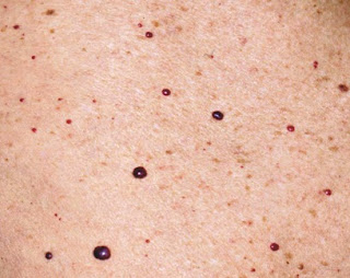
Cherry Hemangiomas (Campbell De Morgan spots Or Senile Angiomas)


Cherry hemangiomas 
also known as Campbell De Morgan spots or senile angiomas are the most common cutaneous vascular proliferations. They are often widespread and appear as tiny cherry-red papules or macules. Longstanding lesions enlarge slowly over time and take on the appearance of a dome topped with cherry-red to deep-purple papules.

They are more common with advancing age and affect men and women equally.

Clinical Features
  • erythematous, papular lesions
  • typically 1-3 mm in size
  • nonblanching
  • not found on the mucous membranes
Causes: The exact cause of cherry angiomas is unknown, but there may be a genetic factor that

Cellulitis - A Brief Discussion

Cellulitis is a common infection of the skin or subcutaneous tissues with characteristic findings of:
  • erythema with poorly defined borders, 
  • edema, warmth, pain, and limitation of movement.
  • Fever and constitutional symptoms may be present and are commonly associated withm bacteremia. 
     Cellulitis of the right lower extremity characterized by sharply demarcated erythema an edema.


Predisposing factors include:


  • trauma, 
  • lymphatic or venous stasis, 
  • immunodeficiency (including diabetes mellitus), and 
  • foreign bodies.

Common etiologic organisms include:

  • group A β-hemolytic Streptococcus 
  • Staphylococcus aureus in nonintertriginous skin, and  gram-negative organisms or mixed flora in intertriginous skin and ulcerations. 
  • In immunocompromised hosts, Escherichia coli, Klebsiella species, Enterobacter species, and Pseudomonas aeruginosa are common. 
  • In recent years, there has been a dramatic increase in the incidence of community- acquire methicillin-resistant S aureus (CA-MRSA), particularly in cellulitis associate with a cutaneous abscess.