Friday, September 30, 2016

A Case Of Pemphigoid Gestationis


A 37-year-old pregnant woman presented to the hospital with severe preeclampsia. After all medical methods were tried and failed to control her severe preeclampsia, a joint decision was made to induce
labor to save the life of the mother. The pregnancy was too early for the fetus to survive.
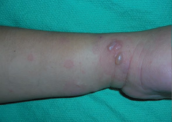
The following day she began to develop the lesions shown in picture above. Her past history includes antiphospholipid syndrome with multiple pregnancy losses. This was her third episode of having such skin lesions.
A diagnosis of pemphigoid gestationis was made after biopsy results from the lesions.

Case Discussion
 Pemphigoid Gestationis

Pemphigoid gestationis is a rare autoimmune bullous dermatosis of pregnancy. The disease was originally known as herpes gestationis because of it visual similarities to herpes simplex infection. However, that term has fallen out of favor because pemphigoid gestationis is not associated with active or prior herpes virus infection.

Pemphigoid gestationis, is defined as a bullous or blistering disease that is associated with pregnancy or with trophoblastic tumors

Pathology: 

• The pathophysiology of the disease involves immunoglobulin (Ig) G antibodies that attack cells in the skin.

Thursday, September 29, 2016

A 70-Year-old Woman Presents with Pain and Swelling in the Joints of Both Hands

A 70-year-old woman presents with pain and swelling in the joints of both hands, which impedes her normal activities. Her pain is better in the morning after resting and worse after she has been working with her hands. She denies stiffness. On examination, you find bony enlargement of some distal interphalangeal (DIP) and proximal interphalangeal (PIP) joints on both hands ( shown in picture above) Radiographs confirm the presence of Heberden and Bouchard nodes. She begins taking 1 g of acetaminophen twice a day and has significant improvement in her pain and function.

What is the diagnosis?

Osteoarthritis.

What is the differential diagnosis for a patient presenting with musculoskeltal pain ? 

Musculoskeletal pain can also be caused by:

A Case Of Urticaria (Hives)


A 45 Year old woman presents to her doctor complaining of itching and red patches on her skin of the legs, arms and abdomen. It first appeared 2 weeks back and has been increasing since then. She recalls having similar symptoms in the past at least 4 times . She relates it to start whenever she drinks citrus fruit juice. She was prescribed antihistamines and her symptoms gradually improved. She was advised to avoid food that trigger the allergy.

Case Discussion:

Hives also known as Urticaria is a skin reaction that presents as a red, raised, itchy skin rash. It appears and fades repeatedly. It usually happens due to an allergy.
Chronic hives is defined when the symptoms last more than six weeks or recur over months or years.

Pathophysiology; Whenever there is an allergic reaction triggered by a substance-allergen , body releases histamine . Histamine causes tiny blood vessels t leak fluid which accumulates in the skin causing the rash and swelling.

Causes : Females and young adults are more prone to utricaria. Each individual may develop symptoms due to a particular allergen to which his/her body responds and produces histamine.
Some of the common causes include:

Wednesday, September 28, 2016

Modified Duke criteria for Infective Endocarditis

Which of the following is/are included in the major criteria of the M

A) Positive blood cultures.
B) Janeway lesions (painless macules on palms and
soles).
C) Echocardiographic evidence of valvular vegetation.
D) A and B.
E) A and C.


The correct answer is “E.” The modified Duke criteria were developed to provide clinicians with standardized criteria for the diagnosis of endocarditis. They have been validated by pathologic examination and are more sensitive than other endocarditis criteria systems.

Interpretation Of PPD Skin Testing (Tuberculin Skin Test)


An 80 Year old female living in nursing home comes to her doctor for the interpretation of her tuberculosis (TB) skin test. 5-tuberculin unit injection of purified protein derivative (PPD) was interpreted as 0 mm diameter of induration after 48 hours.

What should be the next step?

A) Repeat the PPD now.
B) Repeat the PPD in 2 weeks.
C) Repeat the PPD in 1 year.
D) Declare the patient free of TB.
E) Obtain a chest x-ray.

Answer And Discussion: Discussion:

Sunday, September 25, 2016

A 25 Year Old Man Presents With Cough, Low grade fever And Night sweats For 3 Weeks


A 25 -year-old man from the third world country presents to his doctor with a persistent cough for 3 weeks, low grade fever, and night sweats. His chest x-ray  is shown above. The X ray shows mediastinal and right hilar lymphadenopathy and right upper lobe consolidation concerning
for primary tuberculosis (shown by arrows).
The patient's sputum is sent for acid-fast bacillus (AFB) stain and cultures, and the results show acid-fast bacilli consistent with Mycobacterium species.
While culture results are pending, the patient is started on 4 antituberculosis drugs. Fortunately, the sputum  culture result shows pan susceptible Mycobacterium tuberculosis, and his treatment is continued.

Case Discussion:

Tuberculosis:
Introduction: Tuberculosis (TB) is a bacterial infection caused by Mycobacterium . tuberculosis, an
obligate intracellular pathogen that is aerobic, acid fast, and non encapsulated.
TB primarily involves the lungs, although other organs are involved in one-third of cases.

More than 8 million cases occur annually around the world, with nearly 2 million TB-related deaths.  95% of TB deaths occur in low- and middle-income countries.

Risk Factors: Risk factors for infection or progression to active TB include:

• Populations who are at risk to overcrowding and malnutrition.
• HIV
• Immunocompromised states (e.g., cancer, treatment with tumor necrosis factor antagonists)
• Chronic diseases such as diabetes mellitus  or chronic renal failure/hemodialysis
• Malignancy.
• Genetic susceptibility.
• Injection drug users
• Smoking
• Personnel who work or live in high-risk settings (e.g., prisons, long-term care facilities, and hospitals).
• Adult women (ratio 2:1 adult man).
• Older age (both infection and progression).
• Children younger than 4 years of age who are exposed to high-risk individuals.
• Recent infection

Clinical Features: 

  • The disease may be asymptomatic in some patients.
  • TB can affect any organ system.

  • Manifestation of active TB can be classified as pulmonary or extrapulmonary.
  • Saturday, September 24, 2016

    Effect Of Hyperuricaemia On Kidney.

    The above picture shows uric acid kidney stone kept in the pathology lab. Let us briefly study the effects of Hyperuricaemia on kidney.

    Causes Of Hyperuricaemia: High levels of urate in blood is known as hyperuricaemia. It may result from increased turnover (15%) or reduced excretion of urate (85%)
    The causes are given below:

    • Drugs: cytotoxics, thiazides, loop diuretics,pyrazinamide
    • Increased cell turn over: Lymphoma, leukemia, psoariasisf, hemolysis, muscle death-rhabdomyolysis. 
    • Reduced excretion: Promary gout, chronic kidney disease, lead nephropathy, hyper parathyroidism, pre eclampsia
    • Others; Hypertension, Hyperlipedaemia 
    • Lesch-Nyhan syndrome - a disorder of purine synthesis
    Hyperuricaemia and Renal Failure: Severe renal failure from any cause may be associated with hyperuricaemia, and rarely this may cause gout. Sometimes the relationship of cause and effect is reversed so that it is hyperuricaemia that causes the renal failure. This can occur in cytotoxic treatment (tumor lysis syndrome) and in muscle necrosis.

    How Urate Causes Renal Failure?
    Urate is poorly soluble in water so over excretion can lead to crystal precipitation. Renal failure occurs most commonly becauseurate precipitates in the renal tubules. This may occur at a plasma level > 1.19mmol/L. In some instances uretric obstruction from urate crystals may occur. This responds to retrograde uretric catheterization and lavage.

    Thursday, September 22, 2016

    A Sucking Blister In A 3 Day Old Baby

    The picture above shows a blister on the of the hand of a 3 day old baby. This baby boy was delivered at full term to a healthy mother with no prenatal complications. Mother mentions that the baby is a vigorous feeder and seems hungry all the time. He has been sucking on his hands and wrists. The baby is otherwise asymptommatic. On examination of the lesion the underlying skin is non erythematous.

    What is the most likely diagnosis?

    A sucking blister

    A Case Of Cervical Spondylosis

    A 55 Year old Man is seen by his general physical with the complains of pain in his neck over the past 6 months. The pain has been gradually increasing but comes and goes with weeks without any pain. He mentions feeling stiffness in his neck early in the morning.
    The pain sometimes spreads to his right shoulder and he feels tingling sensation in his right arm and hand. A Neck X ray was ordered and showed features of Cervical spondylosis:


    Case Discussion

    Cervical Spondylosis: Is an age related degeneration (wear and tear) of the bones (vertebrae) and discs in the neck. It is also known as  cervical osteoarthritis

    According to certain studies more than 85% of people above the age of 60 years have cervical spondylosis but most of these people have no symptoms.

    Etiology and Pathophysiology: As a person ages , the bones and cartilage that make up the backbone and neck gradually starts to wear and tear and develop certain degenerative changes. These changes can include:

    Wednesday, September 21, 2016

    Treatment Of Supraventricular tachycardia in A Hemodynamically Unstable Patient


    A hemodynamically unstable patient is noted to have supraventricular tachycardia. The most appropriate treatment is

    A) Electrical synchronized cardioversion
    B) Carotid massage
    C) Adenosine
    D) Verapamil
    E) Digoxin

    Answer and Discussion
    The answer is A. Electrical synchronized cardioversion

    Supraventricular tachycardia is characterized by a rapid regular rhythm with a narrow QRS complex and abnormal P waves. The heart rate is usually 100 to 200 bpm. Some patients may be asymptomatic; others may experience chest pain, palpitations, and shortness of breath.

    Hemodynamically unstable patients with supraventricular tachycardia require immediate treatment with electrical synchronized cardioversion.

    A Florist Presents With Non Tender Nodule On Her Hand


    A 40-year-old florist presents to your office complaining of a nontender nodule that formed on her hand, then enlarged, and finally ulcerated. In the days that followed, the patient developed similar nodules in the area of the axillary lymphatics. Otherwise she has had no other symptoms.
    The most likely diagnosis is

    A) Cat-scratch fever
    B) Sporotrichosis
    C) Tuberculosis
    D) Blastomycosis
    E) Histoplasmosis

    Answer and Discussion
    The answer is B. Sporotrichosis

    Sporotrichosis is a disease caused by inoculation of the plant Sporothrix schenckii when patients prick themselves with a thorn. The condition is associated with the formation of nodules, ulcers, and abscesses affecting the skin and lymphatic system. Farm workers and those who work around plants (i.e., florists, gardeners, nursery workers, and horticulturists) such as sphagnum moss, rosebushes, and barberry bushes are the most likely to be affected.

    Tuesday, September 20, 2016

    A Case Of Prader willi Syndrome

    A 3 Year old male child is brought to the pediatrician with the major complaint of excessive weight gain and poor intellectual development. Mother mentions that the child was a floppy infant as a baby and had difficulties with feeding but by the time the child reached 2 years of age , he has a very good appetite and constantly craves for food resulting in weight gain. He seems to be hungry all the time. Also his mother has noticed that her son is developmentally slow compared to his elder siblings, His speech was delayed and he started walking just a few months back. At present he has difficulty speaking full sentences and also responds slowly to commands.


    A diagnosis of Prader willi syndrome was made.

    Case Discussion:

    Systemic Conditions Causing Eye Signs:

    Examining the Eye is an important part of general physical examination. The eye can be a host to many diseases and certain systemic disorders can be diagnosed by identifying the specific signs in the eye. A brief review of the systemic conditions causing eye signs is given below:

    1. Granulomatous disorders: Syphilis, Tuberculosis, sarcoidosis, leprosy, brucellosis and toxoplasmosis may inflame either the front chamber (anterior uveitis/iritis) or back chamber. (posterior uveritis/choroiditis).


    2. Systemic inflammatory diseases: may manifest in any part of the eye.

    • iritis in ankylosing spondilytis
    • Reiter's conjunctivitis in Reiter's syndrome
    • Scleritis or episcleritis in rheumatoid arthritis, vasculitis and SLE

    3. Keratoconjuctivitis sicca: is a reduction in tear formation. It causes gritty feeling in the eyes and a

    Monday, September 19, 2016

    Acute Myeloid Leukemia

    Acute Myeloid Leukemia (AML)

    Also known as Acute myelogenous leukemia or Acute non lymphocytic leukemia

    ( The picture shows a blood smear in a patient with AML . Arrows indicate Aeur rods which are diagnostic of AML)

    Introduction: AML is the rapidly progressive cancer of a group of white blood cells called myeloid cells ( which normally develop into various type of mature blood cells like red blood cells, white blood cells and platelets)

    It is the commonest acute leukemia of adults, and its incidence increases with age. AML progresses rapidly and can cause death in few months if not treated.

    Pathophysiology: AML is caused by damage to the DNA of developing cells in the bone marrow. The bone marrow produces immature cells that develop into leukemic white blood cells called myeloblasts. These abnormal cells build up and over crowd the bone marrow disturbing the function of healthy cells. In most cases the cause of theses DNA mutations is unknown.

    Etiology: In most cases it is not known what leads to leukemia but radiation, exposure to certain chemicals and drugs, genetic disorders like Down syndrome and a previous cancer treatment are considered as risk factors for AML

    Acute Lymphoblastic Leukemia

    Acute Lymphoblastic Leukemia: ( ALL)


    Also known as Acute Lymphocytic leukemia or Acute Lymphoid leukemia

    Introduction: ALL is an acute cancer of the white blood cells affecting either B or T lymphocyte cell lines. The term acute comes from the fact that the disease progresses rapidly and creates immature blast cells with bone marrow failure and tissue infiltration.

    Acute Lymphoblastic leukemia ALL is the most common type of cancer in children and luckily it has a good prognosis if treated . It is rare in adults.

    Pathophysiology: In patients with ALL, there is overproduction of lymphobalsts in the bone marrow. These immature blast cells continuously multiply, and cause damage and death by inhibiting the production of normal red blood cells , white blood cells and platelets in the bone marrow. These malignant cells also infiltrate other organs causing damage.

    Etiology: ALL is thought to develop from a combination of genetic susceptibility ( e.g with translocations amd gains and losses of whole chromosome) as well as an environmental trigger.
    Ionizing radiations, eg X rays during pregnancy and Downs syndrome are important associations.

    Clinical Features:
    Patients usually present with recent onset of signs of marrow failure (pallor, fatigue, bleeding, fever, infection). Hepatosplenomegaly and adenopathy are common. males may have testicular enlargement reflecting leukemic involvement. Meningeal involvement may develop later.

    Sunday, September 18, 2016

    A 32 Year Old Man Presents With Unilateral Nasal Obstruction - A Case Of Nasal Polyp

    A 32-year-old man complains of unilateral nasal obstruction for the past several months of gradual onset. On examination of the nose, a nasal polyp is found.


    Case Discussion

    Nasal Polyp:
    Introduction: Nasal polyps are benign lesions arising from the mucosa of the nasal passages including the paranasal sinuses. They are most commonly semitransparent.

    Epidemiology: Prevalence of 1% to 4% of adults; 0.1% of children of all races and classes.
    • The male-to-female ratio in adults is approximately 2:1.
    • Peak age of onset is 20 to 40 years old; rare in children younger than 10 years old.

    Associations: Associated with the following conditions:
    ~ Nonallergic and allergic rhinitis and rhinosinusitis.
    ~ Asthma—In 20% to 50% of patients with polyps.
    ~ Cystic fibrosis.
    ~ Aspirin intolerance—In 8% to 26% of patients with polyps.
    ~ Alcohol intolerance—In 50% of patients with polyps

    Saturday, September 17, 2016

    Studying The Erythrocyte (RBC) Morphology On Blood Smears

    It is important for every medical student and doctor to learn the basic pathology like studying the morphology of normal and abnormal RBC's on blood smears. A brief description is given below.

    Normal RBC Morphology: It is about 7.5 micrometer in diameter, roughly the size of a small lymphocyte. All the cells are of same size and color and have lightly colored area in the center.

    Reticulocytes: are large, grey blue cells admixed with pink (on Wright's stain). Reticulocyte is actually an immature red blood cell without a nucleus. 


    Anisocytosis: variation in RBC size, 
    • Normal MCV is 80-100 fl
    • large cells imply delay in erythroid precursor DNA synthesis caused by folate or B12 deficiency or as a side effect of medication. Macrocytes MCV > 100fl
    • small cells imply a defect in hemoglobin synthesis caused by iron deficiency or abnormal hemoglobin genes. Microcytes MCV < 80 fl 
    RWD - the automated rec cell distribution width is a measure of anisocytosis.
    Anisocytosis is a feature of most anemias


    Poikilocytes: abnormal RBC shapes. They can be of many types and are described below:

    Friday, September 16, 2016

    Description Of Secondary Skin Lesions


    Description Of Secondary Skin Lesions:

    1. Lichenification: A distinctive thickening of the skin that is characterized by accentuated skin fold markings.


    2. Scale: Excessive accumulation of stratum corneum.


    Thursday, September 15, 2016

    Differential Diagnosis Of Foot Ulcer


    The above picture is taken from a 38 year old diabetic man admitted in surgical ward for management of his foot ulcer. 

    How would you approach to a case of Foot Ulcer?

    A foot ulcer is an open sore on the foot. it can be shallow involving only the surface of the skin or deep extending through the full thickness of the skin. It may even involve the tendons, bones and other deep structures.

    Causes: Ulcers on feet are common and may be either vascular or neuropathic in origin. 

    Vascular Causes: 

    1. Large Vessel Disease: 
    • Arteriosclerosis
    • Embolism
    2. Small Vessel Disease:
    • Diabetes
    • Buerger's disease
    • Raynaud's disease
    • Embolism
    • Vasculitis, e.g SLE, rheumatoid arthritis, scleroderma
    Neuropathic Causes:

    1. Peripheral Nerve Lesions:
    • Diabetes
    • Peripheral nerve injuries
    • Leprosy
    • Alcoholism
    2. Spinal Cord Lesions:
    • Spina bifida
    • Tabes dorsalis
    • Syringomyelia
    Neoplastic Causes:
    • Squamaous cell carcinoma
    • Malignant melanoma

    Description Of Primary Skin Lesions


    Description Of Primary Skin Lesions:

    1. Macule: A flat, colored lesion, <2 cm in diameter, not raised above the surface of the surrounding skin. A "freckle" or ephelid, is a prototype pigmented macule.


    2. Patch: A large ( > 2 cm) flat lesion with a color different from the surrounding skin. 


    The difference between a macule and a patch is only in size.


    3. Papule: A small, solid lesion, < 0.5 cm in diameter, raised above the surface of the surrounding skin and hence palpable. ( e.g a closed comedone or  white-head, in acne)

    A 27 Year Old Male Presenting with Nasal Discharge, Congestion and Headache

    A 27 year old male presents to his doctor with symptoms of nasal discharge. nasal congestion and headache which all started about a week ago. He mentions being in a healthy state before these symptoms started and has no significant past history except getting frequent upper respiratory infections which usually resolve on conservative management. He took acetaminophen but is still not getting better.

    Case Discussion:

    Introduction To Upper Respiratory Infections:

    • URIs are among the leading cause of lost of time from work and school.
    • It is difficult to distinguish URIs caused by virus or bacteria since the signs and symptoms are almost the same. 
    • Non specific URIs also known as "the common cold" have no prominent localizing features and usually present with nasal discharge, nasal congestion, cough and sore throat. In more severe cases patients may suffer from hoarseness, fever, malaise and body ache. 
    • The average duration of symptoms is about 1 week. 
    Etiology of URIs: A wide variety of viruses e.g rhinoviruses, coronaviruses, parainfluenza, influenza virus and adenoviruses can cause non specific URIs. Secondary bacterial infections may complicate a few cases and may lead to prolonged course of disease with more severity of symptoms like purulent nasal and throat discharge. 

    Treatment: Symptom based treatment like decongestants, antipyretics and anti inflammatory drugs are all that is required along with conservative measures like warm fluids, saline water gargles, etc.

    Sinus Infections: Infection and inflammation of the sinuses known as Sinusitis is a common reason for visits to primary care physicians. it most commonly involves the maxillary sinus, followed by the ethmoid, frontal and sphenoidal sinus. 


    Acute Sinusitis: