Tuesday, September 18, 2018

The Placenta, Its Membranes, and the Umbilical Cord

The human placenta is a highly sophisticated organ of interface between mother and fetus, often
referred to as the “gate-keeper to the fetus.” Careful examination of the placenta, its membranes,
and the umbilical cord can prove to be a valuable aid in the diagnosis and treatment of the
neonate. Gross examination of the placenta takes five minutes, and more sophisticated examination
should be considered when there is poor pregnancy outcome, recognizable malformations or
abnormalities, multiple gestation, extremes of amniotic fluid volume, severe intrauterine growth
retardation, short umbilical cord (< 32 cm), and profound acidemia.
The maternal surface of the placenta (decidual plate) is soft, spongy and dark red; and the fetal surface (chorionic plate) is shiny and steel blue to gray.
The placenta, membranes, and umbilical cord weigh approximately 400 to 600 g at birth.

Abnormalities in structure can result in an inefficient transport of oxygen and nutrients to the developing baby. Despite this importance, it is one of the least understood and investigated human organs.

A succenturiate (accessory) lobe is common and has no effect on the fetus. This occurs in about 3 to 5% of deliveries.  Its importance arises from the fact that it may be retained within the uterus and cause postpartum bleeding.


Fetal surface of a bipartite or bilobed placenta (placenta duplex). The two parts of the placenta
are of nearly equal size and this occurs in about 1% of deliveries. Note that the lobes are separated by membranes. The umbilical cord may insert into one or other lobe, or may insert between the two.

In a circumvallate (circummarginate) placenta the fetal surface may be reduced if decidual tissue has made its way between the amnion and chorion. This appears as a yellow, peripheral, hyalinized fold circumscribing the edge of the chorionic plate. This type of placenta has been reported to be a cause of antepartum bleeding and premature labor


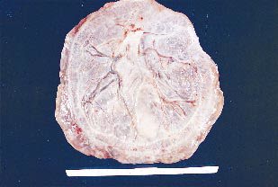
This is an example of placenta membranacea (placenta diffusa). These placentas are rare. The ovum implants too deeply, the villae of the chorion fail to regress, and the placental tissue develops over the entire surface of the chorion. The placenta is very thin and is associated with poor fetal growth and antepartum hemorrhage. There may be previa type bleeding.

Friday, November 3, 2017

Fungal Infection of Nails

Fungal infection of the nails has been classified into four types :

1. White superficial onychomycosis presents with diffuse or speckled white discoloration of the surface of the toenails. It is caused primarily by Trichophyton mentagrophytes, which invades the nail plate. The organism may be scraped off the nail plate with a blade, but treatment is best accomplished by the addition of a topical azole antifungal agent.


2. Distal subungual onychomycosis presents with foci of onycholysis under the distal nail plate or along the lateral nail groove, followed by development of hyperkeratosis and yellow-brown discoloration. The process extends proximally, resulting in nail plate thicken and separation from the nail bed. Trichophyton rubrum and, occasionally, T. mentagrophytes infect the toenails; fingernail disease is almost exclusively due to T. rubrum, which may be associated with superficial scaling of the plantar surface of the feet and often of one hand. These dermatophytes are found most readily at the most proximal area of the nail bed or adjacent ventral portion of the nail plates that are involved. Topical therapies such as ciclopirox 8% lacquer may be effective for solitary nail infection. Because of their long half-life in the nail, terbinafine or itraconazole may be effective when given as pulse therapy (1 wk of each mo for 3–4 mo). Either agent is superior to griseofulvin, fluconazole, orketoconazole. The risks, the most concerning of which is hepatic toxicity, and costs of oral therapy must be weighed carefully against the benefits of treatment for a condition that generally causes only cosmetic problems.

Wednesday, November 1, 2017

Screening and Diagnosis of Celiac Diseases

Screening for celiac disease has been recommended for specific risk factors.

The anti-endomysium IgA antibody and anti-tissue transglutaminase IgA antibody tests are highly sensitive and specific in identifying individuals with celiac disease.

The anti-endomysium IgA antibody test is an immunofluorescent technique and is relatively expensive; interpretation is operator dependent and prone to errors so that it has largely been replaced by anti-tissue transglutaminase IgA antibody tests, which are simpler to perform and have similar sensitivity and specificity.

Anti-gliadin IgA and IgG and anti-reticulin IgA antibody tests are no longer recommended tests due to lack of specificity.

The anti-endomysium IgA and anti-tissue transglutaminase IgA antibody test can be falsely negative with IgA deficiency, which is associated with an increased incidence of celiac disease. Measurement of serum IgA concentration is mandatory to assure that false-negative results in IgA-deficient individuals are excluded. If celiac disease is suspected in patients with IgA deficiency, intestinal biopsy may be required. Because screening with antibodies may identify patients without documented celiac disease on biopsy, it is important to set the lower limit of antibody titers high enough to avoid false-positive results.

A. Normal
B. Celiac disease


Small Intestinal Biopsy.

Definitive diagnosis of celiac disease requires small intestinal biopsy, as none of the available serologic tests are 100% reliable. The characteristic histologic changes include partial or total villous atrophy, crypt elongation and decreased villous/crypt ratio, increased number of intraepithelial lymphocytes, intraepithelial lymphocyte mitotic index >0.2%, decreased height of epithelial cells, and loss of nuclear polarity. The mucosal involvement can be patchy, so multiple biopsies must be obtained.

Monday, October 30, 2017

Some Limb Deformities in a Newborn

1. Metatarsus adductus. In metatarsus adductus, the forefoot is deviated medially and is slightly supinated. In the normal foot, a line drawn through the hindfoot will pass between the second and third toe. With metatarsus adductus, this line will pass lateral to the third toe. The prognosis is excellent, with most cases of metatarsus adductus resolving spontaneously.


2. Calcaneovalgus. Calcaneovalgus deformity of the foot is commonly associated with lateral tibial torsion. The forefoot is abducted, and the ankle is severely dorsiflexed to where the foot folds against the anterolateral surface of the tibia. The deformity is usually flexible, and the foot can be passively placed in the normal position. Calcaneovalgus deformity of the foot usually resolves spontaneously.


3. Talipes equinovarus. Talipes equinovarus, or club foot, has an incidence of approximately 1.5 in 1,000. Fifty percent of the time, the condition is bilateral. It can be associated with other conditions such as spina bifida and arthrogryposis. The foot turns inward and downward and remains tight in this position. Talipes equinovarus requires immediate evaluation since the timing of corrective casting, if necessary, can affect optimal outcome.

Tuesday, August 22, 2017

A 55 Year Old Man Presents To Emergency With Chest Pain For 6 Hours...

The ECG shown below was recorded in the A & E department from a 55-year-old man who had had chest pain at rest for 6 hours. There were no abnormal physical findings.
What does the trace show, and how would you manage him?


The ECG shows:
• Sinus rhythm
• Normal axis
• Normal QRS complexes
• ST segment depression - horizontal in leads V3-V4, downward-sloping in leads I, VL, V5-V6

Monday, July 31, 2017

Important Clinical Findings In The Tympanic Membrane and Middle Ear

“Chalk” patches. White areas of tympanosclerosis are common findings on examination of the ear drum. They are of little significance in themselves, and the hearing is often normal.

A past history of otorrhea in childhood or grommet insertion is usual. Chalk patches do occur with no
apparent past otitis media. Extensive tympanosclerosis with a rigid drum is a sequela of past otitis media, and the ossicles, too, may be fixed or noncontinuous.

Scarring of the drum. A gossamer-thin membrane can be seen to close this previously well-defined central perforation (arrow).


At first sight with the auriscope, a central perforation would appear to be the diagnosis; more careful
examination with a pneumatic otoscope will show that this thin membrane moves and seals the defect, giving reassurance that the drum is intact.

Scarring of the drum with retraction onto the round window, promontory, and incus is also evidence of past otitis media.

It is sometimes difficult to be sure whether this type of drum is intact; a thin layer of epithelium indrawn onto the middle-ear structures may seal the middle ear, and examination with the operating
microscope may be necessary to be certain of an intact drum.

Scarred tympanic membrane. A scarred tympanic membrane in which the drum has become atelectatic and indrawn onto the long process of the incus and promontory.


A retracted tympanic membrane which is thin and indrawn onto the long process of the incus (a), head of the stapes (b), promontary (c), and round window (d).

The stapedius tendon is also seen in this panoramic view obtained with a fiberoptic endoscope.

Creatinine Kinase (CK-MB) Learning Flash Card


Monday, July 17, 2017

Onychomycosis and Diabetes: Patient Case Challenges

Onychomycosis is a common infection that can lead to potentially severe complications in patients who have impaired circulation, peripheral neuropathy, or are immunocompromised. This infection is particularly common in patients with diabetes, many of whom have one or more of these conditions. Onychomycosis treatment reduces risks, but is complicated by the potential for drug-drug interactions between systemic antifungals and the large number of medications often taken by these patients. Several topical therapies are available, including two newer agents that became available in 2014, increasing the potential for individualized treatment selection.

The following case-based activity will test your ability to select appropriate therapies in patients with type 2 diabetes and onychomycosis, while bringing you up to date on risks and management of toenail infections in this vulnerable patient population.

Case :
Albert is a 55-year-old man with type 2 diabetes who presents to your office with thickened, elongated, yellowed toenails (picture shown below). He reports that he first noticed the changes in his toenails about 3 months ago, at which time most of his nails were already involved. Albert was diagnosed with type 2 diabetes 4 years ago, and his blood sugar is currently poorly controlled on metformin alone. He has elevated triglyceride levels for which he is taking a low dose of simvastatin. He is clinically obese, which may have contributed to the fact that he did not notice his toenail infection until it was well entrenched.


On clinical examination, you observe that all of Albert’s toenails are yellow in color and 6 have white lateral streaks. Two nails show involvement extending proximally to the lunula. Several nails, including both great toenails, show subungual hyperkeratosis that is greater than 2 mm thick and two are onycholytic. You estimate that at least 75% of the overall area of Albert’s toenails is infected, with involvement seen in all 10 toenails.

Which of the following clinical characteristics is a risk factor for onychomycosis in patients with diabetes?
A. Female sex
B. Neuropathy
C. Renal dysfunction
D. Younger age

Answer: B. Neuropathy
Discussion: Patients with diabetes can develop peripheral neuropathy, which contributes to the risk of developing onychomycosis, perhaps because this condition impedes proper foot care and allows unrecognized trauma to the nail. 
Men with diabetes are approximately 3 times more likely to develop onychomycosis compared with women with diabetes, and the prevalence of onychomycosis increases with age. Onychomycosis does not seem to be linked to the presence or absence of renal dysfunction in patients with diabetes.

Onychomycosis and Diabetes
Onychomycosis is highly prevalent in patients with diabetes. A North American study of 550 consecutive patients with diabetes found abnormal-appearing toenails in 46% of patients and mycological evidence of onychomycosis in 26% of patients. After adjusting for age, the researchers calculated a 34.9% prevalence in the overall US diabetes population, an estimate that is similar in other studies. Onychomycosis was 2.77 times more likely in patients with diabetes compared with individuals without diabetes. Prevalence in the one-third of patients with type 1 diabetes in this study was lower but still elevated compared with individuals without diabetes, with an odds ratio of 1.69. Onychomycosis was also 3 times more likely in men compared with women with diabetes.

Chilaiditi Sign On Chest X ray

The X ray shown below is of an asymptomatic person. 


Description Of Chilaiditi Sign
Chilaiditi described this normal variant in 1911 where the transverse colon is interposed between the right hemidiaphragm and the liver.
Its prevalence is thought to be 0.025%.

Occasional reports describe patients with Chilaiditi’s syndrome where patients complain of intermittent abdominal pain requiring laparotomy to rule out other causes of peritonism, e.g. perforated ulcer, ruptured appendix.

Atrial Fibrillation - ECG and Learning Questions/Answers


ECG Criteria
• P wave: Absent (P may be replaced by fibrillary f wave).
• Rhythm: Irregularly irregular (R-R interval is irregular). (Atrial rate is very high and ventricular rate is less).

According to the rate, atrial fibrillation may be of 2 types:
• Fast atrial fibrillation: Heart rate >100 beats/min.
• Slow atrial fibrillation: Heart rate <100 beats/min.

Q. What is atrial fibrillation?
Ans. It is an arrhythmia where atria beat rapidly, chaotically and ineffectively, while the ventricles respond at irregular intervals, producing the characteristic irregularly irregular pulse. Any conditions causing raised atrial pressure, increased atrial muscle mass, atrial fibrosis, inflammation and infiltration of the atrium can cause atrial fibrillation.

Q. What are the types of atrial fibrillation?
Ans. There are 3 types of atrial fibrillation:
• Paroxysmal: Discrete self-limiting episodes. May be persistent if underlying disease progresses.
• Persistent: Prolonged episode that can be terminated by electrical or chemical cardioversion.
• Permanent: Sinus rhythm cannot be restored.

Wednesday, July 12, 2017

Keloid Scars - A Brief Discussion


Keloid scars are tumor like lesions that arise from the connective tissue of a scar and extend beyond the dimensions of the original wound

Predisposing factors

  • ethnicity: more common in people with dark skin
  • occur more commonly in young adults, rare in the elderly
Pathophysiology: When skin is injured, fibrous tissue, called scar tissue, forms over the wound to repair and protect the injury. In some cases, scar tissue grows excessively, forming smooth, hard growths called keloids. Keloids can be much larger than the original wound.


Common Sites (in order of decreasing frequency):

  • sternum, 
  • shoulder,
  • neck,
  • face, 
  • extensor surface of limbs, 
  • trunk
Keloids should not be confused with hypertrophic scars, which are raised scars that do not grow beyond the boundaries of the original wound.

Tuesday, July 11, 2017

A 26-year-old woman, who has complained of palpitations in the past, presents to the emergency department ....

A 26-year-old woman, who has complained of palpitations in the past, is admitted via the A & E department with palpitations. Her ECG is shown below:


What does the ECG show and what should you do?

The ECG shows:
• Narrow-complex tachycardia, rate about 200/min
• No P waves visible
• Normal axis
• Regular QRS complexes
• Normal QRS complexes, ST segments and T waves

Clinical interpretation
This is a supraventricular tachycardia, and since no P waves are visible this is a junctional, or
atrioventricular nodal, tachycardia.

Diagnosis: Junctional (atrioventricular nodal re-entry) tachycardia.

What to do
Junctional tachycardia is the commonest form of paroxysmal tachycardia in young people, and

A 26 year old man with lesions on his body...

A 26-year-old man is seen for an upper respiratory infection. After removing his shirt, you notice the lesions shown in the picture below. He reports that his father has similar lesions.


The differential diagnosis should include
A) Hypothyroidism
B) Addison’s disease
C) Multiple sclerosis
D) Neurofibromatosis
E) Gardner’s syndrome

Answer:

Ashman Phenomenon - ECG


ECG Findings
• Aberrant ventricular conduction, usually with RBBB pattern.
• Altered durations of the refractory period of the bundle branch or ventricular tissue are present, commonly due to atrial fibrillation, atrial ectopy, and atrial tachycardia

Points to Remember:

1. After depolarization, tissue repolarizes during its refractory period. Refractory period changes with the preceding cardiac cycle, with longer R-R intervals producing longer refractory periods and shorter R-R intervals producing shorter refractory periods.

2. A longer R-R interval lengthens the following refractory period. When an early or premature (ectopic) depolarization reaches the ventricular conduction system before it has completely repolarized, aberrant conduction may occur and be manifest on the ECG with a bundle branch block (BBB) pattern.

3. Ashman phenomenon most commonly appears with an RBBB pattern, since the right bundle has a longer refractory period than the left bundle.

4. Ashman phenomenon is often seen in atrial fibrillation, when a long R-R interval is followed by a much shorter R-R interval.

Pulmonary Alveolar Microlithiasis - Chest X Ray

A 39 years old female came to radiology department for X-ray chest with history of dyspnea on exertion. The X ray is shown as below:


Radiological Findings on the X ray Chest: X-ray chest (in the picture above) shows that the lung fields are diffusely occupied by discrete high density opacities resembling grains of sands. In spite of
superimposition or summations of shadows the individual deposits are identifiable and measure about a 1 mm in diameter thick and streaky. The opacity appears confluent showing the lungs as almost white with obliteration of the mediastinal and diaphragmatic contours and pulmonary vascular marking are indistinct. The density is greater over the lower than the upper zone.

Comments and Explanation: The characteristic finding on the chest radiograph is that of a fine, dense stippling. This is a well-defined, nodular infiltrate which involves both lungs, and may be so extensive as to cause opaque lungs with relative lucency of the mediastinal structures, diaphragm and pleura. There is widespread parenchymal calcification.
In addition to the finding seen, other findings that may be seen include bullae in the lung apices, a zone of increased lucency between lung parenchyma and the ribs (a black pleural line) and pleural calcification.
In some patients concentration of the nodules in subpleural, para septal and peribronchiolar alveoli can produce linear strands of calcification parallel to or perpendicular to the pleural surface.

Bullous Myringitis - Clinical presentation & Management

Bullous myringitis is a direct inflammation and infection of the tympanic membrane secondary to a viral or bacterial agent. Vesicles or bullae filled with blood or serosanguinous fluid on an erythematous tympanic membrane are the hallmarks. Frequently, a concomitant otitis media with effusion is noted.
Typical pathogens are the same as seen in Acute otitis media.

Clinical Presentation: The onset of bullous myringitis is preceded by an upper respiratory tract infection and is heralded by sudden onset of severe ear pain, scant serosanguinous drainage from the ear canal, and frequently some degree of hearing loss.
Otoscopy reveals bullae on either the inner or outer surface of the tympanic membrane.

Bullous Myringitis. A large fluid-filled bulla is seen distorting the surface of the tympanic membrane.

Patients presenting with fever, hearing loss, and purulent drainage are more likely to have concomitant infections, such as Otitis media and otitis externa .

Management: Differentiation between viral and bacterial etiologies for tympanic membrane bullae is not necessary. Although most episodes resolve spontaneously, many physicians prescribe antibiotics. Warm compresses, topical or strong analgesics, and oral decongestants provide symptomatic relief. Referral is not necessary in most cases unless rupture of the bullae is required for pain relief.

Monday, July 10, 2017

The External Ear - Different Conditions With Pictures

Eczematous Otitis Externa.
Eczema of the meatus and pinna (see picture below) may be associated with eczema elsewhere, particularly in the scalp, or it may be an isolated condition affecting only one ear.
Itching is the main symptom, with scanty discharge.


The eczematous type of otitis externa usually settles with the use of a topical corticosteroid and antibiotic drop. Cleaning of the meatus may also be necessary, either with cotton wool on a probe, or
suction and the Zeiss microscope. Otitis externa tends to recur.

A Furuncle In the Meatus
A furuncle in the meatus is the other common type of otitis externa. It is characterized by pain; pain on movement of the pinna or on inserting the auriscope is diagnostic of a furuncle.


Diabetes mellitus must be excluded with recurrent furuncles.

Coronary Angiography - Flash Cards


Monday, July 3, 2017

Foreign Body Seen on X ray

A 65-year-old male presented with cardiogenic shock. He had an emergency CABG (Coronary artery bypass grafting) which was associated with a very stormy peri-operative period.  His CXR (shown in picture below) was taken upon arrival at the Intensive Care Unit (ICU).

What is the most significant abnormality?


Case Discussion:

The CXR shows an opaque density in the region of the right lower zone.
Each lung field on an erect CXR is divided into three zones.

  1. The upper zone is an area which lies above a horizontal line drawn from the medial end of the second rib anteriorly. 
  2. The middle zone lies below this and is bordered inferiorly by a line drawn similarly from the fourth rib. 
  3. The lower zone lies below this. 

This opaque density is similar in configuration to a tooth which was dislodged during emergency
intubation of this patient.

See the X ray again with the arrow pointing to the opacity